. 2005 Nov 15;106(10):3524-31.
doi: 10.1182/blood-2005-03-1243. Epub 2005 Jul 26.

CD4+ T-cell death induced by infectious and noninfectious HIV-1: role of type 1 interferon-dependent, TRAIL/DR5-mediated apoptosis

Affiliations

CD4+ T-cell death induced by infectious and noninfectious HIV-1: role of type 1 interferon-dependent, TRAIL/DR5-mediated apoptosis

Jean-Philippe Herbeuval et al. Blood. .

Abstract

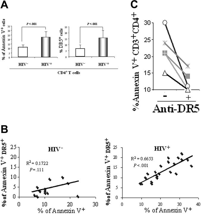
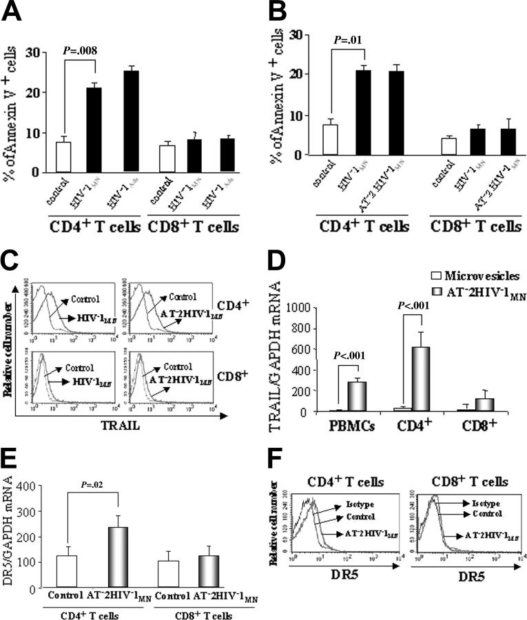
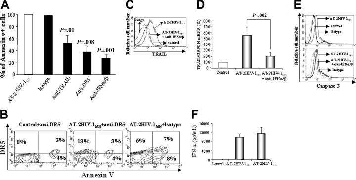
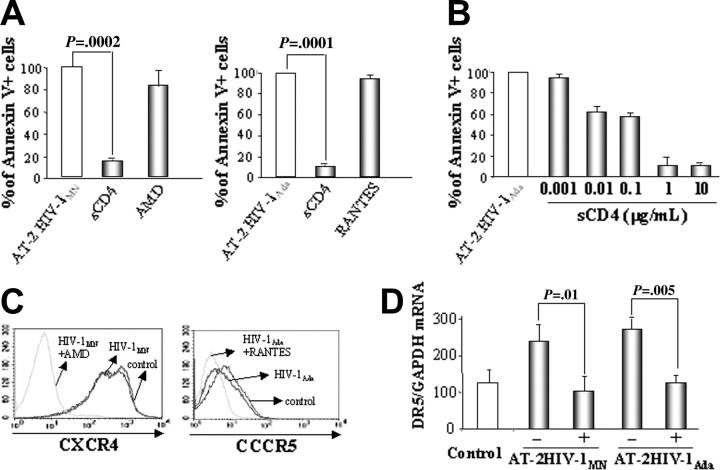
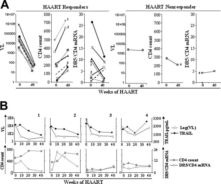
It has been proposed that direct and indirect mechanisms contribute to the unresolved issue of CD4(+) T-cell depletion that results from HIV-1 infection. We recently reported that plasma levels of tumor necrosis factor (TNF)-related apoptosis-inducing ligand (TRAIL) are elevated in HIV-1-infected patients and that they correlate with viral load. The present study investigates the expression of TRAIL death receptor 5 (DR5) in the peripheral-blood mononuclear cells (PBMCs) of HIV-1-infected patients and its role in CD4(+) T-cell death. DR5 expression was elevated and associated with the apoptotic marker annexin V. Apoptosis was reduced in CD4(+) T cells when cultured with anti-DR5 antibody. CD4(+), but not CD8(+), T cells from uninfected donors expressed TRAIL, DR5, and activated caspase-3 when cultured with infectious or noninfectious HIV-1, resulting in preferential apoptosis of CD4(+) T cells. TRAIL, caspase-3 expression, and apoptosis were type 1 interferon (IFN) dependent. Induction of apoptosis and DR5 expression required glycoprotein 120 (gp120)-CD4 interaction. Finally, we analyzed DR5 expression by CD4(+) T cells in highly active antiretroviral therapy (HAART)-treated patients. The decreased viral loads and increased CD4 counts of HAART-responsive patients were associated with a decrease in DR5 mRNA expression by CD4(+) T lymphocytes. We propose a novel model in which a type 1 IFN-regulated TRAIL /DR5 mechanism induces apoptosis of HIV-1-exposed CD4(+) T cells.


Figures

Figure 1.
Figure 1.
Analysis of apoptosis and DR5 in HIV-1–infected patients and uninfected controls. (A) Flow cytometric analysis of annexin V+ and DR5+ CD4+ T cells from 16 uninfected control donors (HIV-) and 22 HIV-1–infected patients (HIV+). P values were calculated using 2-tailed Student t test. (B) Linear regression analysis of DR5-mediated CD4+ T-cell apoptosis (DR5+annexin V+) compared with all apoptotic CD4+ T cells (annexin V+) performed in the same cohort. (C) PBMCs from 5 HIV-1–infected patients were cultured for 48 hours with (+) or without (-) anti-DR5 blocking antibody. CD3+ CD4+ T cells were analyzed for annexin V by FACS.
Figure 2.
Figure 2.
Annexin V, TRAIL, and DR5 expression in T cells. Percentages of annexin V+ cells from HIV-1–uninfected donors determined by flow cytometry in (A) unfractionated PBMCs cultured for 24 hours with microvesicles (control), infectious HIV-1MN, or HIV-1Ada; or (B) isolated CD4+ and CD8+ T cells cultured with microvesicles, or with infectious or AT-2 HIV-1MN. (C) FACS analysis of TRAIL expression on CD4+ and CD8+ T cells cultured for 24 hours with infectious or noninfectious HIV-1MN. (D) TRAIL mRNA expression after 24-hour culture of PBMCs, isolated CD4+ and CD8+ T cells with AT-2 HIV-1MN, or microvesicles. (E) DR5 mRNA expression in CD4+ and CD8+ T cells cultured for 24 hours with AT-2 HIV-1MN or microvesicles. (F) FACS analysis of DR5 expression on CD4+ and CD8+ T cells after 24 hours of culture with AT-2 HIV-1MN (identical results obtained using infectious HIV-1MN; data not shown). Eight percent to 13% of CD4+ T cells expressed DR5, whereas less than 1% of CD8+ T cells did. (A-B,D-E) Mean values with standard errors of 6 independent experiments for each condition tested. (C,F) Representative results of 4 independent experiments. (A-B,E) P values determined by 2-tailed Student t test.
Figure 3.
Figure 3.
Role of type 1 IFN and DR5 in T-cell apoptosis. (A) Effect of monoclonal RIK-2 anti-TRAIL antibody, anti-DR5 antibody, and anti–IFN-α/β antibodies on the percentages of annexin V+ cells resulting from 24-hour cultures of CD4+ T cells with AT-2 HIV-1MN. (B) Two-color flow cytometric analysis of annexin V/DR5 double-positive CD4+ T cells cultured for 24 hours with microvesicles plus anti-DR5 (control), AT-2 HIV-1MN plus isotype, or AT-2 HIV-1 plus anti-DR5 antibody. Control and AT-2 HIV-1 plus anti-DR5–treated cells were then stained using an anti–mouse PE antibody, and cells cultured with AT-2 HIV-1 plus isotype were stained using an anti-DR5 PE antibody. (Top left) Cells are nonapoptotic but DR5+. (Top right) Cells are apoptotic (annexin V+) and DR5+. (C) Effect of IFN-α/β–specific antibodies on TRAIL expression by CD4+ T cells cultured with AT-2 HIV-1MN. (D) Effect of anti–IFN-α/β antibodies on TRAIL mRNA expression induced by culture with AT-2 HIV-1MN for 24 hours. (E) Activated caspase-3 levels in CD4+ T cells cultured for 6 hours with or without AT-2 HIV-1MN (top panel) and in the presence or absence of anti–IFN-α/β antibodies (bottom panel). (F) IFN-α levels, measured by ELISA, in supernatants of isolated pDCs, cultured for 24 hours with microvesicles, AT-2 HIV-1MN, or AT-2HIV-1Ada. (A,D,F) Mean values (± SD) of 6 experiments. (B-C,E) Representative results of 4 independent experiments. (A,D) P values were determined by 2-tailed Student t test.
Figure 4.
Figure 4.
Effect of entry inhibitors on apoptosis and DR5 mRNA expression in T cells. (A) CD4+ cell were cultured for 24 hours with AT-2 HIV-1MN/Ada in the presence of sCD4-IgG (2 μg/mL), AMD-3100 (2 μg/mL), or RANTES (2 μg/mL). Apoptosis was analyzed by flow cytometry using the annexin V method. (B) Titration of sCD4-IgG in blocking AT-2 HIV-1MN–induced apoptosis of CD4+ T cells (μg/mL) (C) Flow cytometric analysis of CXCR4 or CCR5 expression by CD4+ T cells cultured for 24 hours in the presence of AT-2 HIV-1MN with or without AMD-3100 (2 μg/mL) or with or without RANTES (2 μg/mL). (D) DR5 mRNA expression by CD4+ T cells cultured in the presence of HIV-1MN/Ada and with (+) or without (-) sCD4-IgG.
Figure 5.
Figure 5.
Effect of HAART on DR5 mRNA expression in T cells. Eight HIV-1–infected patients started HAART therapy and were followed up for 40 weeks. Samples were collected at weeks 0, 8, 20, and 40. (A) Viral load, CD4 count, and DR5/CD4 mRNA data for weeks 0 and 40 of therapy for 7 HAART responder patients (3 left panels) and 1 HAART nonresponder (3 right panels) patients. (B) Data for weeks 0, 8, 20, and 40 are shown for viral load and plasma TRAIL (top panels) and for CD4 count and DR5/CD4 mRNA ratio (bottom panels) for a representative example (patient 1) of 5 HAART responder patients, the 2 exceptions shown in Figure 4A (second panel) (patients 2 and 3), and the HAART nonresponder (patient 4). Plasma TRAIL levels of patients 1 and 4 were shown in our earlier study. Plasma TRAIL levels of patients 2 and 3 are new data.

References

    1. Brenchley JM, Schacker TW, Ruff LE, et al. CD4+ T cell depletion during all stages of HIV disease occurs predominantly in the gastrointestinal tract. J Exp Med. 2004;200: 749-759. - PMC - PubMed
    1. Mehandru S, Poles MA, Tenner-Racz K, et al. Primary HIV-1 infection is associated with preferential depletion of CD4+ T lymphocytes from effector sites in the gastrointestinal tract. J Exp Med. 2004;200: 761-770. - PMC - PubMed
    1. Groux H, Torpier G, Monte D, Mouton Y, Capron A, Ameisen JC. Activation-induced death by apoptosis in CD4+ T cells from human immunodeficiency virus-infected asymptomatic individuals. J Exp Med. 1992;175: 331-340. - PMC - PubMed
    1. Grossman Z, Meier-Schellersheim M, Sousa AE, Victorino RM, Paul WE. CD4+ T-cell depletion in HIV infection: are we closer to understanding the cause? Nat Med. 2002;8: 319-323. - PubMed
    1. Finkel TH, Tudor-Williams G, Banda NK, et al. Apoptosis occurs predominantly in bystander cells and not in productively infected cells of HIV- and SIV-infected lymph nodes. Nat Med. 1995;1: 129-134. - PubMed

Publication types

MeSH terms