. 2005 Aug 2;102(31):10981-6.
doi: 10.1073/pnas.0504315102. Epub 2005 Jul 26.

NK cytotoxicity against CD4+ T cells during HIV-1 infection: a gp41 peptide induces the expression of an NKp44 ligand

Affiliations

NK cytotoxicity against CD4+ T cells during HIV-1 infection: a gp41 peptide induces the expression of an NKp44 ligand

Vincent Vieillard et al. Proc Natl Acad Sci U S A. .

Abstract

HIV infection leads to a state of chronic immune activation and progressive deterioration in immune function, manifested most recognizably by the progressive depletion of CD4+ T cells. A substantial percentage of natural killer (NK) cells from patients with HIV infection are activated and express the natural cytotoxicity receptor (NCR) NKp44. Here we show that a cellular ligand for NKp44 (NKp44L) is expressed during HIV-1 infection and is correlated with both the progression of CD4+ T cell depletion and the increase of viral load. CD4+ T cells expressing this ligand are highly sensitive to the NK lysis activity mediated by NKp44+ NK cells. The expression of NKp44L is induced by the linear motif NH2-SWSNKS-COOH of the HIV-1 envelope gp41 protein. This highly conserved motif appears critical to the sharp increase in NK lysis of CD4+ T cells from HIV-infected patients. These studies strongly suggest that induction of NKp44L plays a key role in the lysis of CD4+ T cells by activated NK cells in HIV infection and consequently provide a framework for considering how HIV-1 may use NK cell immune surveillance to trigger CD4+ T cells. Understanding this mechanism may help to develop future therapeutic strategies and vaccines against HIV-1 infection.


Figures
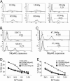
Fig. 1.
Fig. 1.
Identification of 7.1, an anti-NKp44L mAb that specifically inhibits NK lysis mediated by NKp44 on uninfected (UI) and HIV-1-infected (Sf2) U2 cells. (A) U2 cells were incubated with 10 μg/ml NKp30-Ig (30Ig), NKp46-Ig (46Ig), or NKp44-Ig (44Ig) fusion proteins (black lines) or with an Ig control (dotted lines). (B) U2 cells were incubated with 1 μg/ml mAb 7.1 (black line) or with the IgM control (dotted line). (C) mAb 7.1 inhibits NKp44-Ig binding. HIV-1-infected U2 cells that had previously been stained with either 10 μg of NKp44-Ig fusion protein (black line in Lower) or control NKp46-Ig fusion protein (black line in Upper) were incubated with mAb 7.1 (gray line) or IgM control (dotted line). The percentage of cells expressing NKp44L, after NCR-Ig incubation, is noted. (D) Inhibition of NK lysis of U2 cells treated with mAb 7.1 by purified Il-2-activated NK cells. (E) Inhibition of NK lysis of U2 cells by purified IL2-activated NK cells, incubated with 10 μg/ml of anti-NKp44 mAb. UI, uninfected cells; Sf2, HIV-infected cells. These results are representative of at least three independent experiments.
Fig. 2.
Fig. 2.
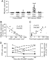
Expression of NKp44L in CD4+ T cells from HIV-infected individuals is associated with disease stage. (A) Specific expression of NKp44L on CD4+ T cells in HIV-infected patients stained with mAb 7.1. The horizontal lines mark the mean value. NKp44L expression is reported as percentage of positive cells. (B) Inverse correlation of NKp44L expression in CD4+ T cells with peripheral blood CD4 cell count in HIV-infected patients. (C) Correlation between NKp44L expression in CD4+ T cells and viral load. (D) Time course of CD4 cell count (open symbols) and NKp44L expression (filled symbols) in three independent subjects (triangle, square, and circle) on highly active antiretroviral therapy.
Fig. 3.
Fig. 3.
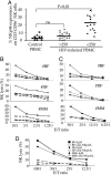
High sensitivity to NK lysis of CD4+ T cells expressing NKp44L. (A) Expression of NKp44 on CD3-CD56+NK cells from healthy donors and HIV-infected patients with a CD4 cells count >250 or <250 per mm3. The percentage of CD3-CD56+NK cells that expressed NKp44 was reported. ns, nonsignificant. Cytotoxic activity of NK92 NK cell line (B) and IL2-activated autologous NK cells (C) was analyzed against CD4+ T cells that did (square) or did not (circle) express NKp44L from three HIV-infected patients (patients BF, 250 CD4+ cells per mm3; DF, 182 CD4+ cells per mm3; MM, 213 CD4+ cells per mm3). Cytotoxic activity was analyzed after treatment with anti-NKp44 mAb. Filled circles, CD4+NKp44L- T cells plus IgM isotype; open circles, CD4+NKp44L- T cells plus anti-NKp44 mAb. Filled squares, CD4+NKp44L+ cells plus IgM isotype; open squares, CD4+NKp44L+ T cells plus anti-NKp44 mAb. (D) Inhibition of cytotoxicity of fresh autologous NK cells without IL2-activation by anti-NKp44L mAb.
Fig. 4.
Fig. 4.
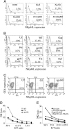
NKp44L is specifically induced by HIV-1. (A) Expression of NKp44L after incubation with AT-2 inactivated HIV-1 particles. CD4+ T cells stained with anti-NKp44L mAb (thick gray line) or IgM isotype control (thin black line). (B) Expression of NKp44L after infection with recombinant vaccinia virus expressing gp160 or gp41 HIV-1 proteins. CD4+ T cells were infected with recombinant vaccinia viruses expressing HIV protein, and then stained with anti-NKp44L mAb (thick gray line) or with IgM isotype control (thin black line). UI, uninfected cells; WT, CD4+ T cells infected with a wild type vaccinia virus. Treatment with recombinant gp160 protein induces NKp44L expression (C) and NK lysis (D). CD4+ T cells were incubated with 5 μg/ml control protein (Ctl), or a recombinant gp160 protein (rgp160) or without protein (UT). The percentage of CD4+ T cells expressing NKp44L was noted for each panel. Lysis activity was performed by using IL2-activated autologous NK cells. (E) Inhibition of gp160 induction in presence of anti-NKp44L mAb. The results are representative of at least three independent experiments.
Fig. 5.
Fig. 5.
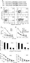
Critical role of the SWSNKS motif. (A) Sequences of gp41-C146 (WT) and two mutated control peptides (Ctl1 and Ctl2). (B) Induction of NKp44L expression. CD4+ T cells were treated with 5 μg/ml peptide and then were stained with no. 7.1 anti-NKp44L mAb. The percentage of CD4+ T cells expressing NKp44L is noted for each panel. (C) Sensitivity of CD4+ T cells incubated with these peptides to IL2-activated autologous NK cells. (D) Inhibition of NK lysis by anti-NKp44L mAb. Effects of NKp44L were inhibited with an anti-gp41-C146 peptide polyclonal antibody. Purified CD4+ T cells from two highly HIV-1 infected patients (CG, 322 CD4+ cells per mm3 and 148,100 HIV copies per ml; and BT, 208 CD4+ cells per mm3 and 255,300 HIV copies per ml) were cultured 2 days in presence of 10 units/ml IL-2, and then treated overnight with several concentrations of anti-gp41-C146 Abs. (E) Inhibition of NKp44L expression in presence of anti-gp41-C146 antibody. (F) Inhibition of NK lysis in presence of anti-gp41-C146 Abs. Cytotoxic activity was performed by using IL2-activated autologous NK cells. Open circles, untreated cells; filled squares, triangles, and diamonds, CD4+ T cells treated with 1, 10, and 20 μg/ml of anti-gp41-C146 Abs, respectively. The results are representative of at least four independent experiments.

References

    1. Flodstrom, M., Shi, F. D., Sarvetnick, N. & Ljunggren, H. G. (2002) Scand. J. Immunol. 55, 432-441. - PubMed
    1. Moretta, L. & Moretta, A. (2004) EMBO J. 23, 255-259. - PMC - PubMed
    1. Biassoni, R., Cantoni, C., Pende, D., Sivori, S., Parolini, S., Vitale, M., Bottino, C. & Moretta, A. (2001) Immunol. Rev. 181, 203-214. - PubMed
    1. Scalzo, A. A. (2002) Trends Microbiol. 10, 470-474. - PubMed
    1. Reyburn, H. T., Mandelboim, O., Vales-Gomez, M., Davis, D. M., Pazmany, L. & Strominger, J. L. (1997) Nature 386, 514-517. - PubMed

Publication types

MeSH terms