Review
. 2005 Aug;14(8):1945-54.
doi: 10.1110/ps.051417505.

Remodeling protein complexes: insights from the AAA+ unfoldase ClpX and Mu transposase

Affiliations
Review

Remodeling protein complexes: insights from the AAA+ unfoldase ClpX and Mu transposase

Briana M Burton et al. Protein Sci. 2005 Aug.

Abstract

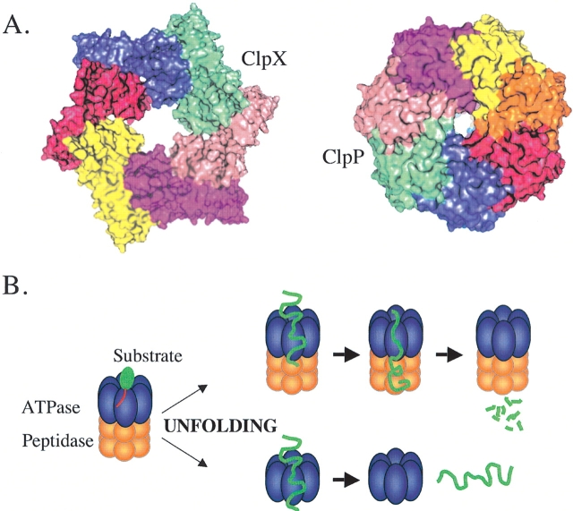
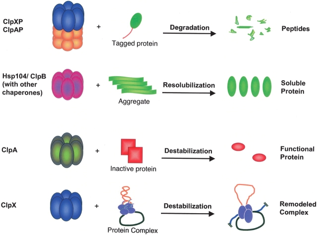
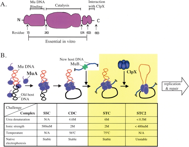
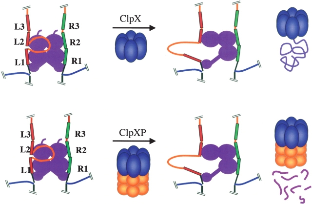
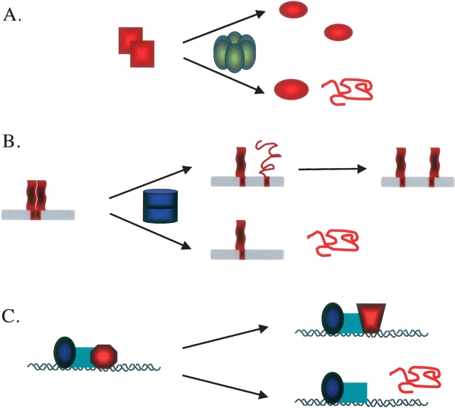
Multiprotein complexes in the cell are dynamic entities that are constantly undergoing changes in subunit composition and conformation to carry out their functions. The protein-DNA complex that promotes recombination of the bacteriophage Mu is a prime example of a complex that must undergo specific changes to carry out its function. The Clp/Hsp100 family of AAA+ ATPases plays a critical role in mediating such changes. The Clp/Hsp100 unfolding enzymes have been extensively studied for the roles they play in protein degradation. However, degradation is not the only fate for proteins that come in contact with the ATP-dependent unfolding enzymes. The Clp/Hsp100 enzymes induce structural changes in their substrates. These structural changes, which we refer to as "remodeling", ultimately change the biological activity of the substrate. These biological changes include activation, inactivation (not associated with degradation), and relocation within the cell. Analysis of the interaction between Escherichia coli ClpX unfoldase and the Mu recombination complex, has provided molecular insight into the mechanisms of protein remodeling. We discuss the key mechanistic features of the remodeling reactions promoted by ClpX and possible implications of these findings for other biological reactions.


Figures

Figure 1.
Figure 1.
Clp/Hsp100 proteins. (A) ClpX and ClpP structure. The Clp/Hsp100 ATPases form hexameric ring structures out of six identical subunits (ClpX shown left). Spacefill representations of ClpX and ClpP demonstrate that ClpP also has rotational symmetry, although sevenfold, and a central axial pore. To form the ClpXP protease the two protein assemblies align to form a stacked barrel with a central channel (the details of how the symmetry mismatch is accommodated in the complex are unknown). The regulatory ATPases thus flank the ends of the peptidase (bound to one or both ends), regulating which proteins may gain access to the peptidase. (B) Degradation and unfolding pathways for ClpX/ClpXP. Unfolding is initiated by binding of substrate to ClpX; binding occurs via a recognition tag (shown in red). Then, in a reaction requiring many rounds of ATP hydrolysis by ClpX, the substrate protein is unfolded. The unfolding reaction is thought to occur while the polypeptide is being pulled through the central pore of ClpX (enzyme subunits are shown as transparent, to show the substrate threading through). This unfolded chain can then be translocated into the ClpP chamber (top pathway) or released into solution if ClpX is not associated with ClpP (bottom pathway).
Figure 2.
Figure 2.
Remodeling reactions. Remodeling is defined as a change in the biological activity of the substrate proteins usually by alteration of the structure of those proteins. (Top) Degradation as catalyzed by ClpXP or ClpAP. The remaining reactions are schematics of the resolublization of heat-induced aggregates by ClpB or Hsp104, and two nonproteolytic remodeling reactions: the conversion of RepA dimers into monomers by ClpA, and the destablization of the Mu transposase/DNA complex by ClpX.
Figure 3.
Figure 3.
Mu transpososomes and the requirement for ClpX. (A) Domain structure of Mu transposase, MuA. The N-terminal domain of transposase is responsible for most of the DNA binding, including recognition of the sequences at the ends of the Mu genome. The large middle domain contains the conserved acidic amino acids that form the active site responsible for both the DNA cleavage and joining reactions. In the C-terminal domain, the last 10 residues of the protein are required for recognition by ClpX. The minimal portion of the protein required for recombination is also denoted. (B) Mu recombination. Monomers of MuA transposase assemble into the stable synaptic complex (SSC) by binding site-specifically to the ends of the Mu DNA. The transposase cleaves one strand at each end of the Mu DNA, to make the cleaved donor complex (CDC). The CDC is then poised to join the exposed 3′ hydroxyl groups of the Mu DNA to a new host DNA molecule. An activator protein, MuB, delivers the target DNA molecule to the transposase. Finally, the transposase catalyzes the DNA joining reaction to produce the strand transfer complex (STC). The table outlines the different stabilities of transpososomes to various challenges. Note that the STC is the most stable complex. After Mu recombination is complete, the transposase remains stably associated with the recombination joints as an STC. The ATP-dependent activity of ClpX, in the absence of ClpP, is necessary and sufficient to destabilize the STC, forming a “fragile” complex, or STC2. The fragile complex, which still has transposase associated with the DNA, is defined by its sensitivity to numerous treatments resisted by the STC. The fragile complex is required for recruitment of bacterial replication machinery to the recombination site and therefore for replicating the Mu DNA.
Figure 4.
Figure 4.
Selective destabilization: model for ClpX-mediated Mu transpososome remodeling. MuA binding sites L1–3 and R1–3 are depicted. Work described in this review defines the following important elements for ClpX-mediated remodeling of Mu transpososomes. First, ClpX uses its unfolding activity when it interacts with transposase subunits in the strand transfer complex. Second, only one subunit or a small subset of subunits is actually contacted by ClpX, and therefore, only a subset is released from the complex into solution. Finally, some physical characteristic of the complex, perhaps an inherent asymmetry, may dictate which subunits are unfolded by ClpX. Thus, the remodeled complex has a preferred configuration in which specific deprotection of DNA in and around the L1 binding site is observed. This region of newly accessible DNA might serve as loading site for the host replication machinery. By this model, the same fragile complex could be generated by either ClpX or ClpXP. Figure reprinted with permission from Elsevier (Burton and Baker 2003).
Figure 5.
Figure 5.
Remodeling other protein complexes by selective destabilization. (A) RepA activation by ClpA. In vitro ClpA can activate phage P1 replication initiator protein RepA. The inactive dimeric form of RepA must be converted to monomers that bind to the origin DNA, oriP1, with high affinity. Following the selective destabilization model, ClpA could unfold one of the subunits from the dimer, thus releasing the other subunit for DNA binding. This unfolding could be partial, allowing the contacted subunit to resume its native conformation and also be active for oriP1 binding (top pathway). Instead, the contacted subunit could be completely and irreversibly unfolded, which might, in turn, lead to degradation of the contacted RepA subunit (bottom pathway). The structure of RepA is unknown; thus, it is unclear the extent to which unfolding of one subunit would disrupt the structure of the second subunit in the dimer. (B) NSF-catalyzed SNARE recycling. SNARE proteins are recycled to allow for multiple rounds of membrane fusion by the help of the NSF complex. In this case, NSF might dissociate the SNARE complexes merely by breaking interactions in the soluble domain, depicted here as unfolding of the coiledcoil domains (top pathway). Alternatively, NSF could completely extract a SNARE subunit from the lipid bilayer when unfolding that subunit (bottom pathway). (C) AAA+-mediated activation of transcription. Proteasomal AAA subunits that promote transcriptional elongation may function via selective destabilization of assembled transcription complexes at certain promoters. The proteasomal ATPases could either promote a conformational change in a specific subunit necessary for elongation (top pathway), or alter the complex by unfolding and removing an inhibitory component (bottom pathway).

References

    1. Aldaz, H., Schuster, E., and Baker, T.A. 1996. The interwoven architecture of the Mu transposase couples DNA synapsis to catalysis. Cell 85 257–269. - PubMed
    1. Baker, T.A. and Mizuuchi, K. 1992. DNA-promoted assembly of the active tetramer of the Mu transposase. Genes & Dev. 6 2221–2232. - PubMed
    1. Bochtler, M., Hartmann, C., Song, H.K., Bourenkov, G.P., Bartunik, H.D., and Huber, R. 2000. The structures of HsIU and the ATP-dependent protease HsIU-HsIV. Nature 403 800–805. - PubMed
    1. Burton, B.M. and Baker, T.A. 2003. Mu transpososome architecture ensures that unfolding by ClpX or proteolysis by ClpXP remodels but does not destroy the complex. Chem. Biol. 10 463–72. - PubMed
    1. Burton, B.M., Williams, T.L., and Baker, T.A. 2001. ClpX-mediated remodeling of mu transpososomes: Selective unfolding of subunits destabilizes the entire complex. Mol. Cell 8 449–454. - PubMed

MeSH terms

LinkOut - more resources