. 2005 Dec;53(12):1511-6.
doi: 10.1369/jhc.5A6704.2005. Epub 2005 Jul 26.

Detection of myelination using a novel histological probe

Affiliations

Detection of myelination using a novel histological probe

Zhongmin Xiang et al. J Histochem Cytochem. 2005 Dec.

Abstract

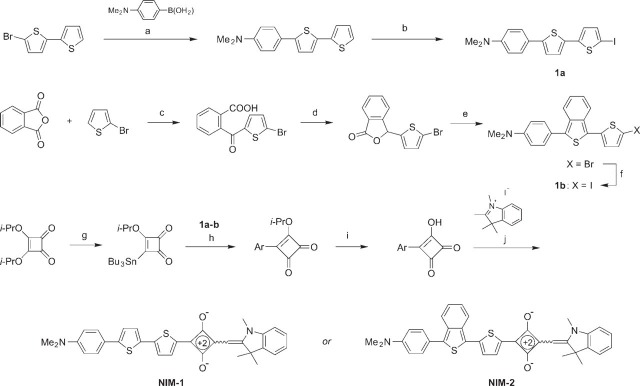
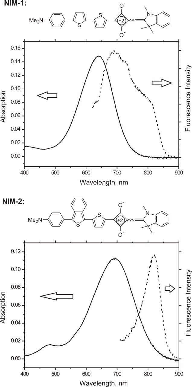
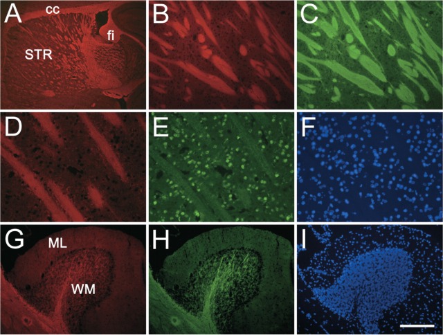
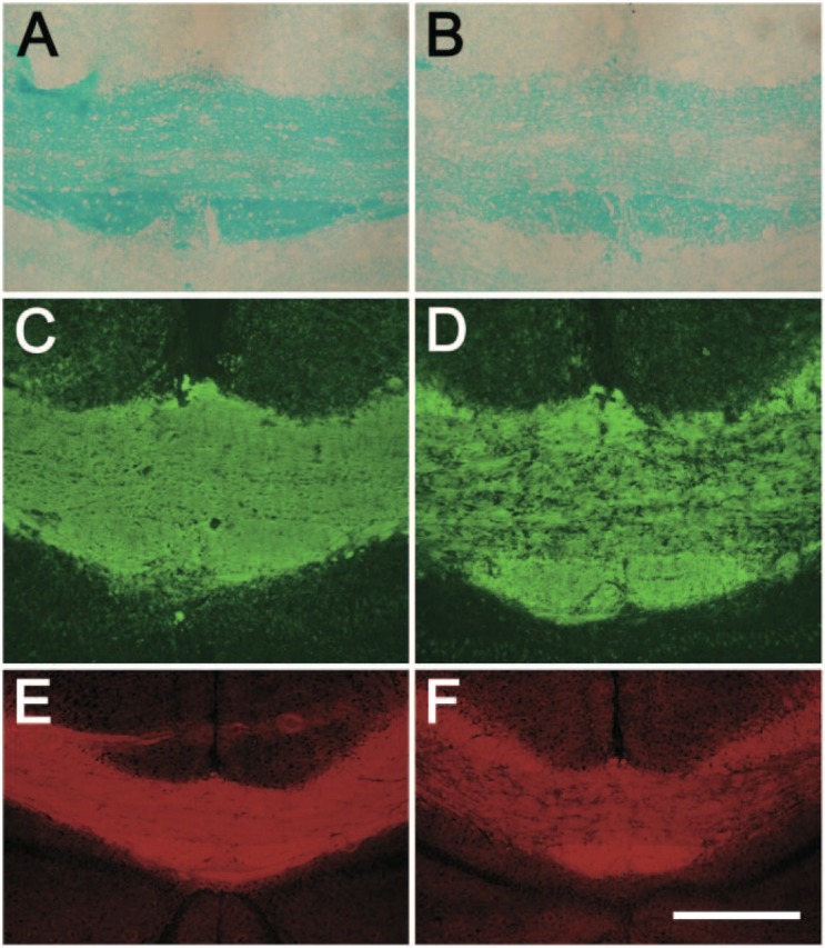
Current methods for myelin staining in tissue sections include both histological and immunohistochemical techniques. Fluorescence immunohistochemistry, which uses antibodies against myelin components such as myelin basic protein, is often used because of the convenience for multiple labeling. To facilitate studies on myelin, this paper describes a quick and easy method for direct myelin staining in rodent and human tissues using novel near-infrared myelin (NIM) dyes that are comparable to other well-characterized histochemical reagents. The near-infrared fluorescence spectra of these probes allow fluorescent staining of tissue sections in multiple channels using visible light fluorophores commonly used in immunocytochemistry. These dyes have been used successfully to detect normal myelin structure and myelin loss in a mouse model of demyelination disease.


Figures

Figure 1.
Figure 1.
Synthesis of NIM-1 and NIM-2 dyes. Reaction conditions: (a) Pd(PPh3)4 (catalyst), K2CO3, toluene-EtOH-H2O, 85C, 66%; (b) 1) n-BuLi, TMEDA, THF, −70C, 2) I2, −70C to 20C, 75% in two steps; (c) AlCl3, CH2Cl2, 40C, 47%; (d) NaBH4, NaHCO3, H2O, 20C, 75%; (e) 1) 4-Me2N-C6H4-MgBr, THF, 0C to 20C; 2) Lawesson reagent, CH2Cl2, 20C, 58% in two steps; (f) 1) n-BuLi, THF, −70C; 2) I2, −70C to 20C, 75% in two steps; (g) n-Bu3Sn-SiMe3, n-Bu4N+CN- (catalyst), THF, 66%; (h) Pd(PPh3)4 (catalyst), CuI (catalyst), DMF, 60C; (a) 63%; (b) 91%; (i) aqueous HCl, THF, 20C; (a) 83%; (b) 91%; (j) quinoline (catalyst), n-BuOH-C6H6, reflux; (a) 55%; (b) 42%. Abbreviations: TMEDA, N, N, N', N'-tetramethylethylenediamine; THF, tetrahydrofuran; DMF, N, N-dimethylformamide; Cul, copper iodide; Ph, phenyl; Bu, butyl; Me, methyl; Et, ethyl; Pr, propyl; PPh3, triphenylphospine. Ar refers to the bithienyl residue either from 1a (for NIM-1) or from 1b (for NIM-2). The exact stereochemistry of the ethylene double bonds in the structures of the dyes was not determined and is indicated by a squiggly line.
Figure 2.
Figure 2.
Structure and absorption/emission spectrum of NIM dyes. NIM-1 and NIM-2 were solubilized in DMSO at 2.1 μM (1.1 μg/ml) and 2.1 μM (1.2 μg/ml), respectively. Absorption maxima: NIM-1 641 nm (× = 72,000); NIM-2 692 nm (× = 54,000).
Figure 3.
Figure 3.
NIM-1 staining in the normal mouse brain. Para-sagittal sections were stained with NIM-1 (red, A, B, D, G) costained with either myelin basic protein (MBP) (green, C, H) or NeuN (green, E), and mounted with the nuclear stain DAPI (blue, F, I). NIM-1 stains distinct white matter structures in the brain (A), such as striatum (STR), corpus callosum (cc), and fimbria (fi). In the striatum (B-F), NIM-1 stains fiber bundles (B), completely overlapping MBP staining (C). The compatibility of NIM-1 staining (D) with other fluorescence staining is further demonstrated by NeuN immunostaining (E) and nuclear stain DAPI (F). In the cerebellum folium, NIM-1 stains the white matter (WM), colocalizes with MBP (H), while leaving molecular layer (ML) unstained. Bars: A = 500 μm; B, C = 100 μm; D-F = 50 μm; G-I = 200 μm.
Figure 4.
Figure 4.
NIM-1 efficiently detects demyelination in a cuprizone-induced animal demyelination model. Brain coronal sections from control mice (A, C, E) and cuprizone-fed mice (B, D, F) were stained with Luxol Fast Blue (A, B), MBP (C, D), and NIM-1 dye (E, F). Shown are middle portions of CC. Bars: A-D = 100 μm; E-F = 200 μm.

References

    1. Hirano M, Osakada K, Nohira H, Miyashita A. (2002) Crystal and solution structures of photochromic spirobenzothiopyran. First full characterization of the meta-stable colored species. J Org Chem 67: 533–540 - PubMed
    1. Klüver H, Barrera E. (1953) A method for the combined staining of cells and fibers in the central nervous system. J Neuropathol Exp Neurol 12: 400–403 - PubMed
    1. Kuhn H, Schweig A. (1967) Theoretical treatment of solvent effects on the electronic spectra of polar organic dye molecules. Chem Phys Lett 1: 255–258
    1. Matsushima GK, Morell P. (2001) The neurotoxicant, cuprizone, as a model to study demyelination and remyelination in the central nervous system. Brain Pathol 11: 107–116 - PMC - PubMed
    1. Nakazumi H, Colyer C, Kaihara K, Yagi S, Hyodo Y. (2003) Red luminescent squarylium dyes for noncovalent HAS labeling. Chem Lett 32: 804–805

Publication types

Substances