. 2005 Dec 1;392(Pt 2):335-44.
doi: 10.1042/BJ20050465.

STAT3-mediated transcription of Bcl-2, Mcl-1 and c-IAP2 prevents apoptosis in polyamine-depleted cells

Affiliations

STAT3-mediated transcription of Bcl-2, Mcl-1 and c-IAP2 prevents apoptosis in polyamine-depleted cells

Sujoy Bhattacharya et al. Biochem J. .

Abstract

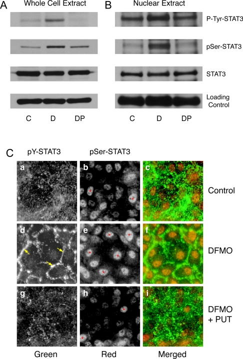
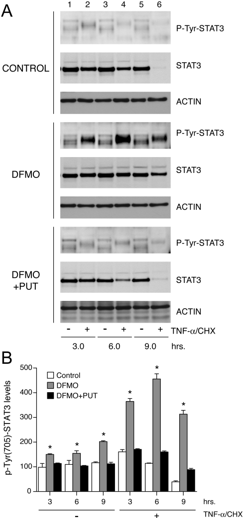
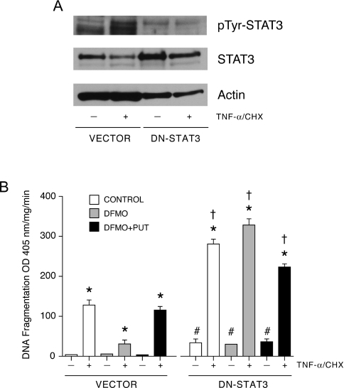
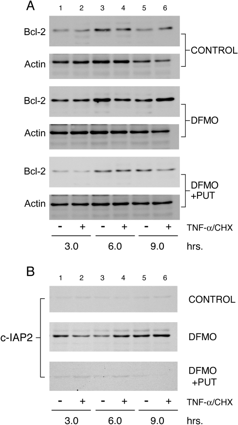
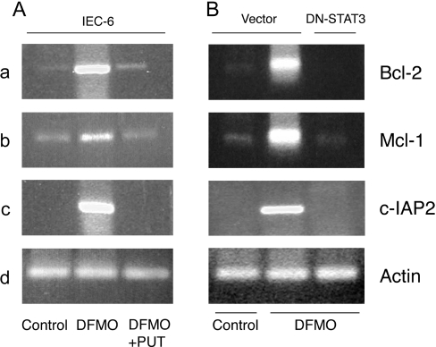
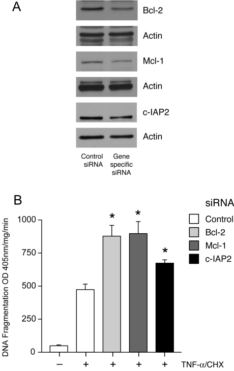
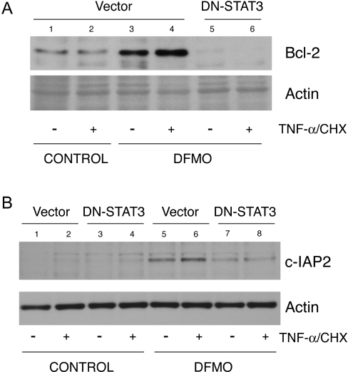
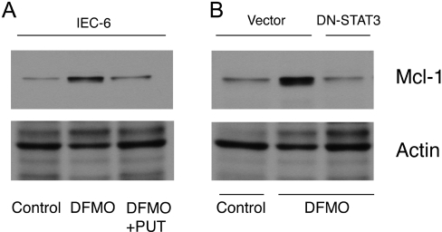
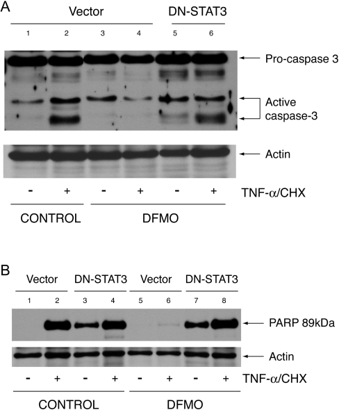
Activation of STAT3 (signal transducer and activator of transcription 3) plays a crucial role in cell survival and proliferation. The aim of the present study was to clarify the role of STAT3 signalling in the protection of polyamine-depleted intestinal epithelial cells against TNF-alpha (tumour necrosis factor-alpha)-induced apoptosis. Polyamine depletion by DFMO (alpha-difluoromethylornithine) caused phosphorylation of STAT3 at Tyr-705 and Ser-727. Phospho-Tyr-705 STAT3 was immunolocalized at the cell periphery and nucleus, whereas phospho-Ser-727 STAT3 was predominantly detected in the nucleus of polyamine-depleted cells. Sustained phosphorylation of STAT3 at tyrosine residues was observed in polyamine-depleted cells after exposure to TNF-alpha. Inhibition of STAT3 activation by AG490 or cell-membrane-permeant inhibitory peptide (PpYLKTK; where pY represents phospho-Tyr) increased the sensitivity of polyamine-depleted cells to apoptosis. Expression of DN-STAT3 (dominant negative-STAT3) completely eliminated the protective effect of DFMO against TNF-alpha-induced apoptosis. Polyamine depletion increased mRNA and protein levels for Bcl-2, Mcl-1 (myeloid cell leukaemia-1) and c-IAP2 (inhibitor of apoptosis protein-2). Significantly higher levels of Bcl-2 and c-IAP2 proteins were observed in polyamine-depleted cells before and after 9 h of TNF-alpha treatment. Inhibition of STAT3 by AG490 and DN-STAT3 decreased Bcl-2 promoter activity. DN-STAT3 decreased mRNA and protein levels for Bcl-2, Mcl-1 and c-IAP2 in polyamine-depleted cells. siRNA (small interfering RNA)-mediated inhibition of Bcl-2, Mcl-1 and c-IAP2 protein levels increased TNF-alpha-induced apoptosis. DN-STAT3 induced the activation of caspase-3 and PARP [poly(ADP-ribose) polymerase] cleavage in polyamine-depleted cells. These results suggest that activation of STAT3 in response to polyamine depletion increases the transcription and subsequent expression of anti-apoptotic Bcl-2 and IAP family proteins and thereby promotes survival of cells against TNF-alpha-induced apoptosis.


Figures

Figure 1
Figure 1. STAT3 phosphorylation in polyamine-depleted cells
(A) Western blot analysis of whole cell extracts using antibodies against phospho-Tyr-705 STAT3 and phospho-Ser-727 STAT3. Blots were stripped and probed for total STAT3 protein, followed by β-actin as an internal loading control. (B) Western blot analysis of nuclear extracts probed with phospho-Tyr-705 and phospho-Ser-727 STAT3 antibodies. Membranes were stained with Ponceau-S as a loading control. IEC-6 cells were grown in DMEM/5% (w/v) dFBS (C) with or without 5 mM DFMO (D) or DFMO plus 10 μM putrescine (DP) for 3 days and serum-deprived for 24 h. Representative blots from three observations are shown. (C) Cells were grown on poly(L-lysine)-coated coverslips for 3 days and serum-deprived for 24 h. PUT, putrescine. Cells were stained using phospho-Tyr-705 and phospho-Ser-727 STAT3-specific antibodies followed by a secondary antibody conjugated to either Alexa-Fluor 488 (green) or Alexa-Fluor 568 (red). STAT3 phosphorylation at Tyr-705 (green) and Ser-727 (red) was visualized using a UV fluorescence microscope. ↓ and * indicate STAT3 localization at the cell periphery and nuclei respectively.
Figure 2
Figure 2. STAT3 activation in response to TNF-α
IEC-6 cells were grown as described in the Experimental section and treated with TNF-α/CHX for 3, 6 and 9 h. (A) Western blot analysis with antibodies against phospho-Tyr-705 STAT3 and total STAT3. Blots were stripped and probed with anti-actin antibody to confirm equal loading of samples. Representative blots from three independent observations are shown. (B) Densitometric analysis of STAT3 activation. *, P<0.005 compared with the respective untreated group. PUT, putrescine.
Figure 3
Figure 3. DN-STAT3 prevents the protective effect of DFMO
(A) Cells transfected with empty vector (vector) or DN-STAT3 were grown as described in the Experimental section and treated with TNF-α/CHX for 3 h. Western blot was performed with phospho-Tyr-705 STAT3-specific antibody. Membrane was stripped and first probed with total STAT3 specific antibody followed by β-actin antibody. (B) DNA fragmentation of vector- and DN-STAT3-expressing cells was determined in response to TNF-α/CHX. *, P<0.005 compared with untreated cells; †, P<0.005 compared with respective TNF-α/CHX-treated vector cells; #, P<0.005 compared with respective untreated vector cells (mean±S.E.M., n=6). PUT, putrescine.
Figure 4
Figure 4. Expression of Bcl-2 and c-IAP2 proteins is elevated in polyamine-depleted cells
IEC-6 cells were grown as described in the Experimental section and treated with TNF-α/CHX for 3, 6 and 9 h. Equal amounts of whole cell extract were separated by SDS/PAGE, and probed with antibodies specific for Bcl-2 (A) and c-IAP2 (B). Membranes were stripped and probed with β-actin antibody to determine equal loading of the samples. PUT, putrescine.
Figure 5
Figure 5. Bcl-2, Mcl-1 and c-IAP2 mRNA levels
(A) IEC-6 cells were grown in DMEM/5% dFBS with or without 5 mM DFMO or DFMO plus 10 μM putrescine (PUT) for 3 days and serum-deprived for 24 h. RNA extraction, reverse transcription and PCR were carried out using gene-specific primers as described in the Experimental section. The PCR products were resolved by agarose gel electrophoresis and stained with ethidium bromide. Representative images from three observations are shown. (B) Vector- and DN-STAT3-transfected cells were grown, as described in the Experimental section for the IEC-6 cells. Total RNA was prepared, reverse-transcribed and PCR was carried out using primers specific for Bcl-2, Mcl-1, c-IAP2 and actin genes as described in the Experimental section. Representative images from three observations are shown.
Figure 6
Figure 6. siRNA-mediated inhibition of Bcl-2, Mcl-1 and c-IAP2 expression and apoptosis
IEC-6 cells were transfected with either control or the respective siRNA as described in the Experimental section. (A) Expression of Bcl-2, Mcl-1 and c-IAP2. Western blot analysis of control and siRNA transfected cell lysates was performed using Bcl-2, Mcl-1 and c-IAP2 specific antibodies. Membranes were stripped and probed for actin as control for sample loading. Representative blots from three observations are shown. (B) DNA fragmentation. Cells were grown in control medium and transfected with either control siRNA or siRNA specific for Bcl-2, Mcl-1 and c-IAP2. After 48 h transfection the cells were serum-starved, and treated with TNF-α/CHX for 3 h and then subjected to DNA fragmentation analysis. Values are means±S.E.M. *P<0.05 compared with control siRNA transfected cells treated with TNF-α/CHX. OD, absorbance.
Figure 7
Figure 7. DN-STAT3 and Bcl-2 and c-IAP2 protein expression
(A) Vector- and DN-STAT3-transfected cells were grown as described in the Experimental section and treated with TNF-α/CHX for 3 h. Lysates were prepared and separated by SDS/PAGE. Proteins were transferred to PVDF membrane and probed with Bcl-2 (A) and c-IAP2 (B) antibodies. Membrane was stripped and probed with β-actin antibody as an internal loading control. Representative blots from three observations are shown.
Figure 8
Figure 8. DN-STAT3 and Mcl-1 protein expression
Parental IEC-6 cells (A) and cells transfected with vector and DN-STAT3 (B) were grown as described in the Experimental section. Lysates were prepared and separated by SDS/PAGE. Proteins were transferred to PVDF membrane and probed with Mcl-1 antibody. Membrane was stripped and probed with β-actin antibody as an internal loading control. Representative blots from three observations are shown.
Figure 9
Figure 9. Caspase-3 activation and PARP cleavage
Cells transfected with vector and DN-STAT3 were grown as described in the Experimental section and treated with TNF-α/CHX or left untreated. Protein (40 μg) from each sample was separated by SDS-gel electrophoresis, transferred to membrane and probed with caspase-3 antibody (A) and a cleaved PARP (Asp-214) rat-specific antibody (B). Membranes were stripped and probed with β-actin antibody as an internal control for equal loading. Representative blots from three experiments are shown.

References

    1. Hall P. A., Coates P. J., Ansari B., Hopwood D. Regulation of cell number in the mammalian gastrointestinal tract–the importance of apoptosis. J. Cell. Sci. 1994;107:3569–3577. - PubMed
    1. Potten C. S. Epithelial cell growth and differentiation 2: Intestinal apoptosis. Am. J. Physiol. Gastrointest. Liver Physiol. 1997;273:G253–G257. - PubMed
    1. Schipper R. G., Penning L. C., Verhofstad A. A. J. Involvement of polyamines in apoptosis. Facts and controversies: effectors or protectors? Semin. Cancer Biol. Rev. 2000;2:55–68. - PubMed
    1. Ray R. M., Viar M. J., Yuan Q., Johnson L. R. Polyamine depletion delays apoptosis of rat intestinal epithelial cells. Am. J. Physiol. Cell Physiol. 2000;278:C480–C489. - PubMed
    1. Tabor C. W., Tabor H. Polyamines. Annu. Rev. Biochem. 1984;53:749–790. - PubMed

Publication types

MeSH terms