. 2005 Oct 7;280(40):34210-7.
doi: 10.1074/jbc.M503523200. Epub 2005 Jul 26.

Mammary serine protease inhibitor (Maspin) binds directly to interferon regulatory factor 6: identification of a novel serpin partnership

Affiliations

Mammary serine protease inhibitor (Maspin) binds directly to interferon regulatory factor 6: identification of a novel serpin partnership

Caleb M Bailey et al. J Biol Chem. .

Abstract

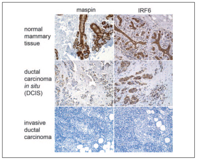
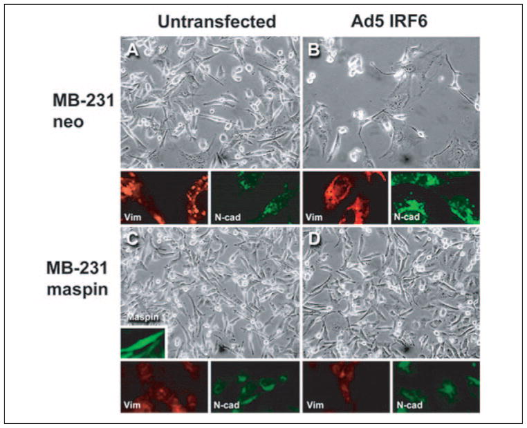
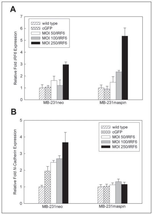
Since its reported discovery in 1994, maspin (mammary serine protease inhibitor) has been characterized as a class II tumor suppressor by its ability to promote apoptosis and inhibit cell invasion. Maspin is highly expressed in normal mammary epithelial cells but reduced or absent in aggressive breast carcinomas. However, despite efforts to characterize the mechanism(s) by which maspin functions as a tumor suppressor, its molecular characterization has remained somewhat elusive. Therefore, in an attempt to identify maspin-interacting proteins and thereby gain insight into the functional pathways of maspin, we employed a maspin-baited yeast two-hybrid system and subsequently identified Interferon Regulatory Factor 6 (IRF6) as a maspin-binding protein. IRF6 belongs to the IRF family of transcription factors, which is best known for its regulation of interferon and interferon-inducible genes following a pathogenic stimulus. Although many of the IRF family members have been well characterized, IRF6 remains poorly understood. We report that IRF6 is expressed in normal mammary epithelial cells and that it directly associates with maspin in a yeast two-hybrid system and in vitro. The interaction occurs via the conserved IRF protein association domain and is regulated by phosphorylation of IRF6. We have shown that, similar to maspin, IRF6 expression is inversely correlated with breast cancer invasiveness. We further demonstrated that the transient re-expression of IRF6 in breast cancer cells results in an increase of N-cadherin and a redistribution of vimentin commensurate with changes in cell morphology, suggestive of an epithelial-to-mesenchymal transition event. Concomitantly, we showed that maspin acts as a negative regulator of this process. These findings help to elucidate the molecular mechanisms of maspin and suggest an interactive role between maspin and IRF6 in regulating cellular phenotype, the loss of which can lead to neoplastic transformation.


Figures

FIGURE 1
FIGURE 1. Amino acid sequence of IRF6
Shading indicates identity (black) or similarity (gray) with other IRFs (IRF3–9). Inverted triangles (▼) indicate sites for introns with the number representing the following exon. The start codon is at the beginning of exon 3, and the stop codon is in exon 9. The functional domains consist of the DNA binding domain (exons 3– 4), a less conserved proline-rich domain (exons 5– 6), the interferon association domain (exons 7– 8), and the serine-rich region (red bar, exon 9). Indicated are a putative nuclear localization sequence (red), the part of IRF6 encoded by the partial cDNA clone pulled out in the original yeast two-hybrid experiment (black box), pentatryptophan residues of DNA binding domain (*), and potential phosphorylation sites (orange).
FIGURE 2
FIGURE 2. Validation of IRF6 antibody (a) and IRF6 and maspin (b– d) expression profile in normal primary and cultured mammary epithelial cells
Post-nuclear cytosolic fractions from 1436N1 cells and Ad5 IRF6-infected MDA-MB-231 cells were evaluated by Western blot (a) before and after preincubation of the antibody with blocking peptides, demonstrating the specificity of the IRF6 antibody. Shown are semiquantitative PCR (b), Western blot analysis (c), and confocal microscopy (d) of IRF6 and maspin in normal primary human mammary epithelial cells (HMEpC) and the immortalized mammary cell line 1436N1. Protein from the cytosolic fraction was evaluated by SDS-PAGE on a 10% gel. Actin was used as a protein loading control. GAPDH was used as an RNA loading control. For confocal microscopy, 1436N1 cells were dual-stained for maspin (green) and IRF6 (red; original magnification ×40).
FIGURE 3
FIGURE 3. a, effect of phosphatase treatment on IRF6 and in vitro confirmation (b– c) of maspin-IRF6 interaction
a, post-nuclear cytosolic fractions from HMEpC cells were treated with lambda-phosphatase (0.4 units) for 1 h at room temperature in the absence of phosphatase inhibitors NaF and sodium orthovanadate. Samples were analyzed using SDS-PAGE and Western blot. b, 500 μg of total protein from the cytosolic fraction of 1436N1 cells was precleared with Protein A-coated Sepharose beads for 30 min and then incubated for 2 h with anti-IRF6 polyclonal antibody at room temperature. Protein A-coated Sepharose beads were added to the lysate and incubated for an additional hour at room temperature. Beads were washed three times in PBS, and then protein was eluted with 2× sample buffer with SDS for 5 min. For phosphatase-treated samples, 500 μg of protein was treated with 2 units of phosphatase for 30 min at room temperature prior to the addition of primary antibody. Lysate was evaluated prior to and following immunoprecipitation and shows the efficient removal of IRF6 from the lysate, whereas only a small portion of maspin was removed. c, full-length IRF6 and eight different IRF6 deletion mutant constructs were expressed as prey and co-transformed with maspin (bait) in the yeast strain AH109. -Leu/-Trp dropout medium was used to select for bait and prey plasmids. -Leu/-Trp/-Ade is a medium stringency dropout medium, and -Leu/-Trp/-Ade/-His is a high stringency dropout medium. The p53-large T antigen interaction was used as a positive control. The graph on the left illustrates the various IRF6 deletion constructs, with arrows depicting the IRF6 fragment used as prey (full-length IRF6 contains exons 3–9). Protein interactions were measured by yeast growth on appropriate selective media.
FIGURE 4
FIGURE 4. IRF6 and maspin expression profiles in poorly invasive and highly invasive metastatic breast cancer lines
Semiquantitative PCR (a) and Western blot analysis (b) of IRF6 and maspin in the poorly invasive neoplastic breast cell lines MCF-7 and T47-D and the highly invasive metastatic cell lines MDA-MB-231 and HS578T. The post-nuclear cytosolic fraction was analyzed. GAPDH was used as an RNA loading control. Actin was used as a protein loading control.
FIGURE 5
FIGURE 5. Immunohistochemical analysis of IRF6 and maspin in normal and neoplastic tissue sections
A comparative analysis of maspin and IRF6 immunoreactivity in archival tissue samples from normal mammary and breast cancer patient tissues with ductal carcinoma in situ (DCIS) and invasive ductal carcinoma. Diaminobenzidine was used as the chromogen.
FIGURE 6
FIGURE 6. Phenotypic analysis of MDA-MB-231 neo and MDA-MB-231 green fluorescent protein-maspin cells following transient infection with Ad5 IRF6
Representative phase-contrast micrographs of MDA-MB-231 neo (MB-231neo) cells untransfected for IRF6 expression (A) and Ad5 IRF6 infected (B) at an m.o.i. of 100 were taken 7 days post-infection. The MDA-MB-231 green fluorescent protein-maspin-transfected cells (MB-231maspin) were either negative for IRF6 expression (C, untransfected) or infected with Ad5 IRF6 (D). Representative samples were also fixed for immunohistochemistry and stained for vimentin (red) and N-cadherin (green) as shown under the representative phase-contrast micrographs. The insert depicts immunofluorescence of MB-231maspin cells. Original magnification was ×20 for all pictures.
FIGURE 7
FIGURE 7. Real-time PCR analysis demonstrating changes in N-cadherin expression following Ad5 IRF6 infection of MDA-MB-231 cells
A, verification of IRF6 expression in MB-231neo and MB-231maspin cells following infection with various concentrations of Ad5 IRF6 (m.o.i. 50, 100, 250). B, changes in N-cadherin expression in the same cells infected with Ad5 IRF6 (m.o.i. 50, 100, 250).

References

    1. Zou Z, Anisowicz A, Hendrix MJC, Thor A, Neveu M, Sheng S, Rafidi K, Seftor E, Sager R. Science. 1994;263:526–529. - PubMed
    1. Sheng S, Carey J, Seftor E, Dias L, Hendrix MJC, Sager R. Proc Natl Acad Sci U S A. 1996;93:11669–11674. - PMC - PubMed
    1. Zhang M, Volpert O, Shi YH, Bouck N. Nat Med. 2000;6:196–199. - PubMed
    1. Jiang N, Meng Y, Zhang S, Mensah-Osman E, Sheng S. Oncogene. 2002;21:4089–4098. - PubMed
    1. Zhang M, Magit D, Botteri F, Shi HY, He K, Li M, Furth P, Sager R. Dev Biol. 1999;215:278–287. - PubMed

Publication types

MeSH terms