. 2005 Jul 26;33(13):4157-63.
doi: 10.1093/nar/gki733. Print 2005.

Induction of transcription within chromosomal DNA loops flanked by MAR elements causes an association of loop DNA with the nuclear matrix

Affiliations

Induction of transcription within chromosomal DNA loops flanked by MAR elements causes an association of loop DNA with the nuclear matrix

Olga V Iarovaia et al. Nucleic Acids Res. .

Abstract

The spatial organization of an approximately 170 kb region of human chromosome 19, including CD22 and GPR40-GPR43 genes, was studied using in situ hybridization of a set of cosmid and PAC probes with nuclear halos prepared from proliferating and differentiated HL60 cells. The whole region under study was found to be looped out into the nuclear halo in proliferating cells. It is likely that the loop observed was attached to the nuclear matrix via MAR elements present at the flanks of the area under study. Upon dimethyl sulfoxide-induced differentiation of the cells the looped fragment became associated with the nuclear matrix. This change in the spatial organization correlated with the activation of transcription of at least two (CD22 and GPR43) genes present within the loop. The data obtained are discussed in the framework of the hypothesis postulating that the spatial organization of chromosomal DNA is maintained via constitutive (basic) and facultative (transcription-related) interactions of the latter with the nuclear matrix.


Figures

Figure 1
Figure 1
A scheme of the area under study. Genes are shown by open rectangles above the scale line. MAR elements are shown by vertical bars. Numeration of MARs is according to ref. (12). Short horizontal bars show positions of two DNA fragments studied in PCR-stop experiments. Insertions of clones used for in situ hybridization experiments are shown below the scale line. The accession numbers of the corresponding DNA sequences deposited in the Gene Bank (AC002132, U62631, AC002511 and AC002997) are presented above the lines showing the insertions.
Figure 2
Figure 2
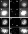
Visualization of the region under study on nuclear halos prepared from non-induced HL60 cells. Panels AD show DAPI staining of nuclear halos and the results of hybridization are shown in panels A′D′. Panels A″D″ show superposition of DAPI staining and hybridization signals, which are seen as black spots over light nuclear halos. Magnification is shown by white bars.
Figure 3
Figure 3
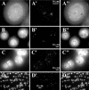
Visualization of the region under study on nuclear halos prepared from DMSO-induced HL60 cells. Panels A–D show DAPI staining of nuclear halos and the results of hybridization are shown in panels A′–D′. Panels A″–D″ show superposition of DAPI staining and hybridization signals, which are seen as black spots over light nuclear halos. Magnification is shown by white bars.
Figure 4
Figure 4
Comparison of the expression of CD22 and GPR43 genes in proliferating (−DMSO) and differentiated (+DMSO) HL60 cells. The figures above the lines loaded with samples show the number of PCR cycles in each case. Each probe was co-amplified with a β-actin fragment to normalize PCR reactions.
Figure 5
Figure 5
Analysis of accessibility of MAR 4 by PCR-stop experiments (A) and a fragment of similar size from the DNA loop (B) for cleavage by endogenous topoisomerase II. Lanes 1–3 were loaded (both in A and B) with DNA samples prepared from control cells (lane 1) and from cells incubated with VM26 (50 μg/ml) for 2 h (line 2) or 5 h (line 3). Lane ‘M’ in panel A was loaded with a molecular weight marker.

References

    1. Paulson J.R., Laemmli U.K. The structure of histone-depleted metaphase chromosomes. Cell. 1977;12:817–828. - PubMed
    1. Hancock R., Hughes M.E. Organization of DNA in the eukaryotic nucleus. Biol. Cell. 1982;44:201–212.
    1. Adolph K.W., Chang S.M., Laemmli U.K. Role of nonhistone proteins in metaphase chromosomes structure. Cell. 1977;12:805–816. - PubMed
    1. Aelen J.M., Opstelsten R.J., Wanka F. Organization of DNA replication in Physarum polycephalum. Attachment of origins of replication and replication forks to the nuclear matrix. Nucleic Acids Res. 1983;11:1181–1195. - PMC - PubMed
    1. Buongiorno-Nardelli M., Gioacchino M., Carri M.T., Marilley M. A relationship between replicon size and supercoiled loop domains in the eukaryotic genome. Nature. 1982;298:100–102. - PubMed

Publication types

Associated data