. 2005 Jul 27;25(30):6939-46.
doi: 10.1523/JNEUROSCI.1674-05.2005.

Programmed and induced phenotype of the hippocampal granule cells

Affiliations

Programmed and induced phenotype of the hippocampal granule cells

Gisela Gómez-Lira et al. J Neurosci. .

Abstract

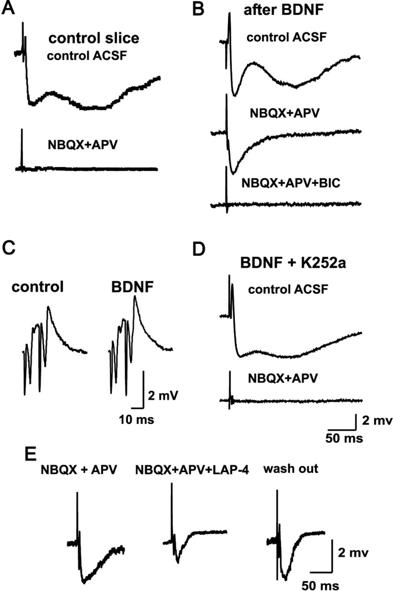
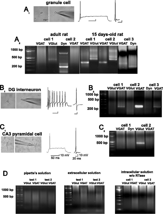
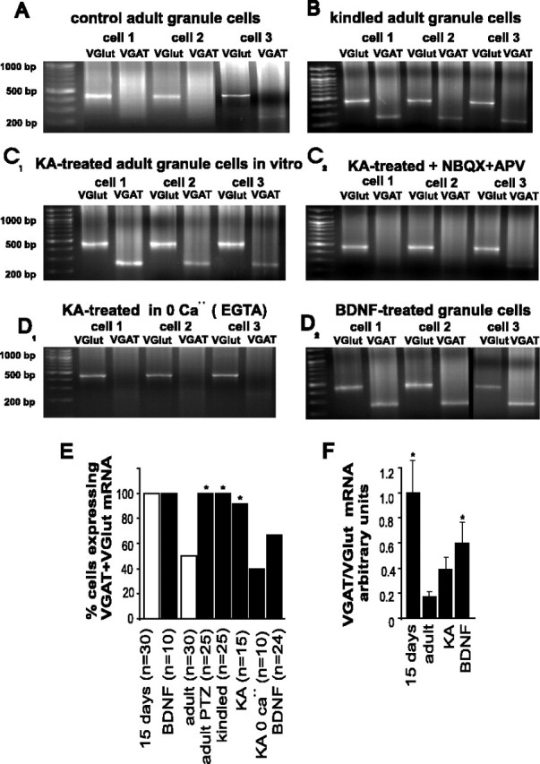
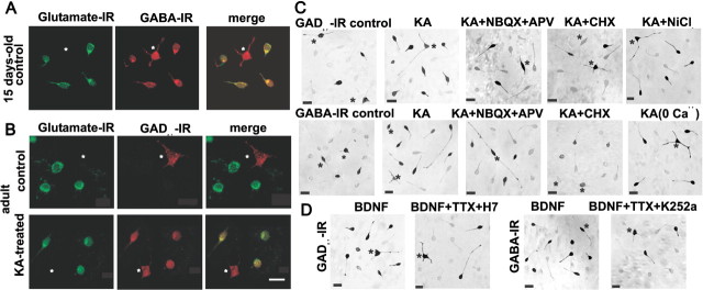
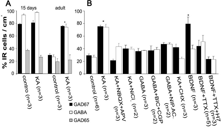
Certain neurons choose the neurotransmitter they use in an activity-dependent manner, and trophic factors are involved in this phenotypic differentiation during development. Developing hippocampal granule cells (GCs) constitutively express the markers of the glutamatergic and GABAergic phenotypes, but when development is completed, the GABAergic phenotype shuts off. With electrophysiological, single-cell reverse transcription-PCR and immunohistological techniques, we show here that short-term (24 h) cultures of fully differentiated adult glutamatergic GCs, which express glutamate, VGlut-1 (vesicular glutamate transporter) mRNA, calbindin, and dynorphin mRNA, can be induced to reexpress the GABAergic markers GABA, GAD67 (glutamate decarboxylase 67 kDa isoform), and VGAT (vesicular GABA transporter) mRNA, by sustained synaptic or direct activation of glutamate receptors and by activation of TrkB (tyrosine receptor kinase B) receptors, with brain-derived neurotrophic factor (BDNF) (30 min). The expression of the GABAergic markers was prevented by the blockade of glutamate receptors and sodium or calcium channels, and by inhibitors of protein kinases and protein synthesis. In hippocampal slices of epileptic rats and in BDNF-treated slices from naive rats, we confirmed the appearance of monosynaptic GABAA receptor-mediated responses to GC stimulation, in the presence of glutamate receptors blockers. Accordingly, GC cultures prepared from these slices showed the coexpression of the glutamatergic and GABAergic markers. Our results demonstrate that the neurotransmitter choice of the GCs, which are unique in terms of their continuing birth and death throughout life, depends on programmed and environmental factors, and this process is neither limited by a critical developmental period nor restricted by their insertion in their natural network.


Figures

Figure 1.
Figure 1.
Mossy fiber monosynaptic GABAergic transmission can be detected when GCs express the markers of the GABAergic phenotype. A, Synaptic responses recorded in CA3 pyramidal cells from naive slices are blocked by the perfusion of the ionotropic glutamate receptors antagonists NBQX and APV. B, In contrast, in preparations exposed to 30 min of BDNF (or to synaptic or direct activation in vivo and in vitro), mossy fiber stimulation provokes bicuculline-sensitive monosynaptic IPSPs in the presence of NBQX and APV. C, BDNF treatment does not induce hyperexcitability, as assessed by field potential responses of CA3 to DG stimulation. D, The Trk receptor inhibitor K252a prevents the effect of BNDF, described in B. E, The pharmacologically isolated monosynaptic GABAergic responses are depressed by l-AP-4, a group III mGluR agonist. All traces are an average of six evoked responses. BIC, Bicuculline; LAP-4, l-AP-4.
Figure 2.
Figure 2.
Developing GCs coexpress VGAT, VGlut-1, and dynorphin (Dyn) mRNAs. Whole-cell recordings in GCs (A1), DG interneurons (B1), and CA3 pyramidal cells (C1) in current-clamp mode permit their identification by the characteristic firing pattern during a depolarizing pulse and by the morphology of their action potentials. A2, Single-cell RT-PCR of two adult GCs shows the presence of VGlut-1 and Dyn mRNAs but not of the VGAT transcript. In contrast, three GCs cultured from 15-d-old rats coexpressed VGlut-1, VGAT, and Dyn mRNAs. B2, Dentate gyrus interneurons expressed VGAT but not Dyn and VGlut-1 mRNAs, whereas CA3 pyramidal cells (C1, C2) expressed VGlut-1 but not VGAT or Dyn mRNAs. D, Single-cell RT-PCR control experiments. The transcripts were undetected after amplification of the contents of the pipette before (left) and after aspiration of extracellular solution (middle) and after amplification of the contents of the cells in the absence of reverse transcriptase (RTase) (right). Calibration for A1 and B1 as in C1.
Figure 3.
Figure 3.
Granule cells cultured from epileptic rats, and from adult naive rats exposed to KA or BDNF, coexpress VGAT and VGlut-1 mRNA. A, Control adult GCs express VGlut-1 and some express barely detectable levels of VGAT mRNA (cell 3). B, In contrast, all GCs cultured from kindled rats coexpress VGlut and VGAT mRNA. C1, KA-treated GC cultures coexpress VGlut-1 and VGAT mRNAs, whereby the expression of the latter is prevented by coincubation of the cells with KA and glutamate receptor blockers (C2) or calcium-free medium (D1). D2, Incubation of adult GC cultures with BDNF produces an upregulation of the expression of VGAT mRNA. E, Summary of the results of the single-cell RT-PCR experiments showing the percentage of adult GCs that coexpressed VGlut-1 and VGAT mRNA under the different experimental conditions. *Significant statistical difference obtained by χ2 test with respect to adult, control at p < 0.05 (n = number of cells). F, Densitometric analysis of VGAT mRNA/VGlut-1 mRNA expression ratio under different experimental conditions. Note that the densitometric measurements included different numbers of cells in each condition, as shown inE. *Significant statistical difference obtained by one-way ANOVA at p < 0.05. Error bars indicate SE.
Figure 4.
Figure 4.
Immunocytochemical determination of glutamate, GAD67, and GABA in cultured GCs. A, Confocal images of short-term cultures of GCs and interneurons (asterisks) of a 15-d-old rat show the coexpression of glutamate-IR and GABA-IR in GCs. B, In cultures from adult rats, GCs express glutamate-IR but not GABAergic markers unless they are exposed to KA, whereby GAD67-IR and glutamate-IR colocalize. C, Immunocytochemical determinations of the GABAergic markers after different treatments. Interneurons can be clearly differentiated and are indicated by asterisks. The number of GCs expressing GAD67 and GABA was increased by the exposure of the cultures to 30 min of KA. The presence of glutamate receptor antagonists, or of Na+ and Ca2+ channel blockers, prevented the expression of the GABAergic markers in the GCs. D, BDNF upregulated the GABAergic markers in GCs, and this effect was prevented by the tyrosine kinase inhibitor with high specificity for the Trk family, K252a. Scale bars: B (for A, B), 15 μm; C, D, 20 μm.
Figure 5.
Figure 5.
Percentage of cultured GCs expressing the GABAergic markers in control conditions and after the different experimental treatments. A, Developing GCs express the GABAergic markers in control conditions and in the presence of KA. Whereas the expression of GAD65 is not modified, GABA and GAD67 are strongly downregulated in adult GCs and upregulated by KA treatment. B, The effects of KA are prevented in the presence of glutamate receptor antagonists and protein synthesis or calcium channel blockers. GABA receptor agonists and antagonists produce no effects. BDNF selectively increased the expression of GAD67. Whereas adding TTX partially prevented the effect of BNDF, the tyrosine kinase inhibitor with high specificity for the Trk family, K252a, and H7, a PKC inhibitor, totally blocked its effect. *Statistically significant difference with respect to the control group at p < 0.05 (ANOVA). Error bars indicate SE.

References

    1. Altar CA, DiStefano PS (1998) Neurotrophin trafficking by anterograde transport. Trends Neurosci 21: 433-437. - PubMed
    1. Ben-Ari Y, Tseeb V, Raggozzino D, Khazipov R, Gaïarsa JL (1994) gamma-Aminobutyric acid (GABA): a fast excitatory transmitter which may regulate the development of hippocampal neurones in early postnatal life. Prog Brain Res 102: 261-273. - PubMed
    1. Bergersen L, Ruiz A, Bjaalie JG, Kullmann DM, Gundersen V (2003) GABA and GABAA receptors at hippocampal mossy fibre synapses. Eur J Neurosci 18: 931-941. - PubMed
    1. Berninger B, Garcia DE, Inagaki N, Hahnel C, Lindholm D (1993) BDNF and NT-3 induce intracellular Ca2+ elevation in hippocampal neurones. NeuroReport 4: 1303-1306. - PubMed
    1. Berzaghi MP, Gutiérrez R, Heinemann U, Lindholm D, Thoenen H (1995) Neurotrophins induce acute transmitter-mediated changes in brain electrical activity. Soc Neurosci Abstr 21: 226.3.

Publication types

MeSH terms

LinkOut - more resources