

Comparison of liquid based cytology and histology for the evaluation of HER-2 status using immunostaining and CISH in breast carcinoma
- PMID: 16049291
- PMCID: PMC1770887
- DOI: 10.1136/jcp.2004.024224
Comparison of liquid based cytology and histology for the evaluation of HER-2 status using immunostaining and CISH in breast carcinoma
Abstract
Background: HER-2 amplification is an important prognostic biomarker and treatment determinant in breast carcinoma.
Aims: To correlate immunocytochemical (ICC) expression of HER-2 and gene amplification determined by chromogenic in situ hybridisation (CISH) using liquid based cytology (LBC) with immunohistochemistry (IHC) and CISH using histological samples of the same breast carcinomas.
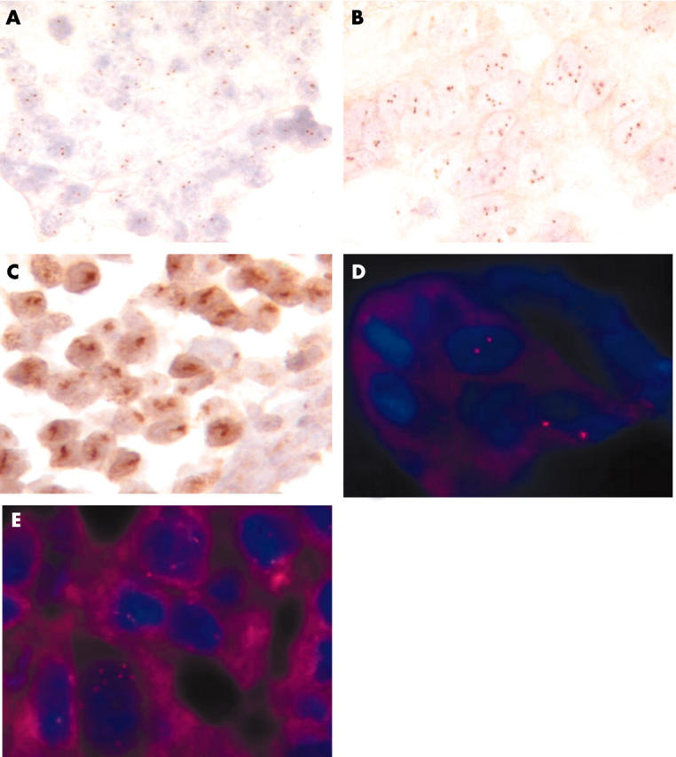
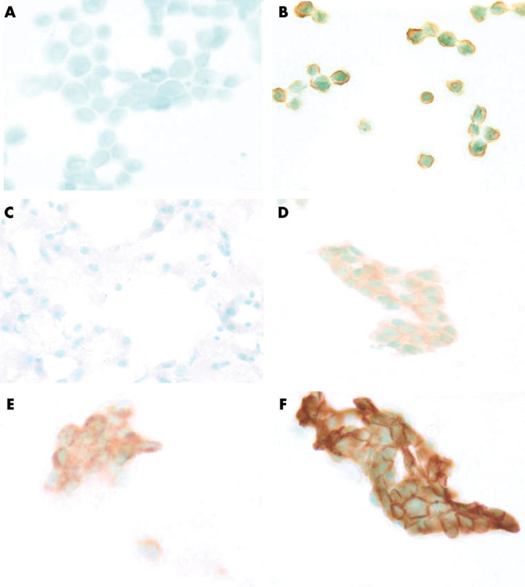
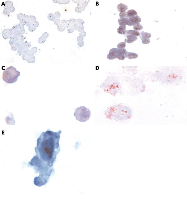
Methods: Frozen sections and cytobrushings of 103 breast carcinomas were analysed. Four techniques were performed on each tumour: two on LBC samples (ICC, and CISH, both graded as positive, indeterminate, or negative) and two on histological samples (IHC and CISH). Two cell lines (MCF-7, negative; BT 474, positive) were used as controls for cytological analysis. A complementary fluorescence in situ hybridisation technique was carried out in histological samples with low amplification (4-10 dots/nucleus).
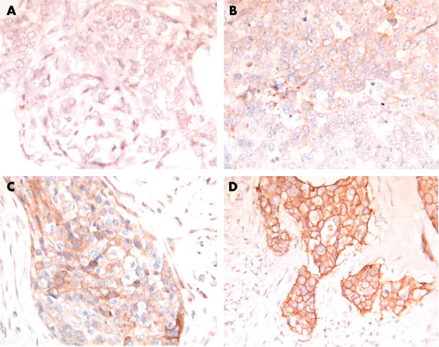
Results: Interobserver agreement for the four techniques calculated by the kappa coefficient indicated a substantial agreement. Nine cases failed in cytology because of poor cellularity. Among 94 cases, 19 were amplified; 73, 12, and 9 tumours were scored 0 or 1+, 2+, and 3+, respectively by IHC and 75, 13, and 6, respectively, by ICC. CISH found no amplification in 72 tumours. Correlations between the IHC and CISH results in the histological and cytological samples were always significant.
Conclusions: Her-2 status could be determined in LBC samples and correlated well with reference histological methods using in situ hybridisation. ICC was less reliable because of the presence of the cytoplasmic membrane. However, these results should be confirmed by a large multicentre study.
Figures
Similar articles
-
HER-2 gene amplification by chromogenic in situ hybridisation (CISH) compared with fluorescence in situ hybridisation (FISH) in breast cancer-A study of two hundred cases.Breast. 2006 Aug;15(4):519-27. doi: 10.1016/j.breast.2005.09.008. Epub 2005 Nov 14. Breast. 2006. PMID: 16290155
-
Evaluation of the clinical significance of HER2 amplification by chromogenic in situ hybridisation in patients with primary breast cancer.Anticancer Res. 2004 Jul-Aug;24(4):2401-6. Anticancer Res. 2004. PMID: 15330190
-
Chromogenic in situ hybridization analysis of HER-2/neu status in cytological samples of breast carcinoma.Cytopathology. 2004 Dec;15(6):315-20. doi: 10.1111/j.1365-2303.2004.00214.x. Cytopathology. 2004. PMID: 15606364
-
Strong correlation between results of fluorescent in situ hybridization and immunohistochemistry for the assessment of the ERBB2 (HER-2/neu) gene status in breast carcinoma.Mod Pathol. 2000 Nov;13(11):1238-43. doi: 10.1038/modpathol.3880228. Mod Pathol. 2000. PMID: 11106082
-
[HER-2 oncogene amplification assessment in invasive breast cancer by dual-color in situ hybridization (dc-CISH): a comparative study with fluorescent in situ hybridization (FISH)].Ann Pathol. 2011 Dec;31(6):472-9. doi: 10.1016/j.annpat.2011.10.013. Epub 2011 Nov 26. Ann Pathol. 2011. PMID: 22172120 Review. French.
Cited by
-
Testing for her2 in breast cancer: current pathology challenges faced in Canada.Curr Oncol. 2012 Dec;19(6):315-23. doi: 10.3747/co.19.1173. Curr Oncol. 2012. PMID: 23300357 Free PMC article.
-
RefCNV: Identification of Gene-Based Copy Number Variants Using Whole Exome Sequencing.Cancer Inform. 2016 Apr 27;15:65-71. doi: 10.4137/CIN.S36612. eCollection 2016. Cancer Inform. 2016. PMID: 27147817 Free PMC article.
-
Chromogenic in situ hybridization compared with other approaches to evaluate HER2/neu status in breast carcinomas.Braz J Med Biol Res. 2013 Mar;46(3):207-16. doi: 10.1590/1414-431x20132483. Epub 2013 Mar 19. Braz J Med Biol Res. 2013. PMID: 23558859 Free PMC article. Review.
-
Bright-field HER2 dual in situ hybridization (DISH) assay on breast cancer cell blocks: a comparative study with histological sections.Breast Cancer. 2016 Nov;23(6):917-921. doi: 10.1007/s12282-015-0664-1. Epub 2016 Jan 8. Breast Cancer. 2016. PMID: 26746842 Free PMC article.
-
Introduction and utility of liquid-based cytology on aspiration biopsy of peripheral nodular lesions of the lung.Oncol Lett. 2014 Mar;7(3):669-673. doi: 10.3892/ol.2013.1763. Epub 2013 Dec 17. Oncol Lett. 2014. PMID: 24520290 Free PMC article.
References
-
- Brandt-Rauf PW, Pincus MR, Carney WP. The c-erb B-2 protein in oncogenesis: molecular structure to molecular epidemiology. Crit Rev Oncog 1994;5:313–29. - PubMed
-
- Coene ED, Schelfhout V, Winkler RA, et al. Amplification units and translocation at chromosome 17q and c-erbB-2 overexpression in the pathogenesis of breast cancer. Virchows Arch 1997;430:365–72. - PubMed
-
- Farabegoli F, Ceccarelli C, Santini D, et al. c-erbB-2 over-expression in amplified and non-amplified breast carcinoma samples. Int J Cancer 1999;84:273–7. - PubMed
-
- Harbeck N, Ross JS, Yurdseven S, et al. HER-2/neu gene amplification by fluorescence in situ hybridization allows risk-group assessment in node-negative breast cancer. Int J Oncol 1999;14:663–71. - PubMed
-
- Slamon DJ, Clark GM, Wong SG, et al. Human breast cancer: correlation of relapse and survival with amplification of the HER-2/neu oncogene. Science 1987;235:177–82. - PubMed
Publication types
MeSH terms
Substances
LinkOut - more resources
Full Text Sources
Other Literature Sources
Medical
Research Materials
Miscellaneous