Review
. 2005 Aug;7(3):317-26.
doi: 10.1016/S1525-1578(10)60560-0.

Experience and strategy for the molecular testing of Duchenne muscular dystrophy

Affiliations
Review

Experience and strategy for the molecular testing of Duchenne muscular dystrophy

Thomas W Prior et al. J Mol Diagn. 2005 Aug.

Abstract

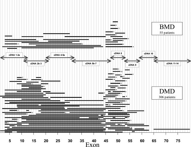
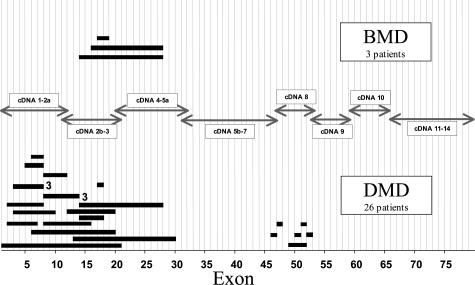
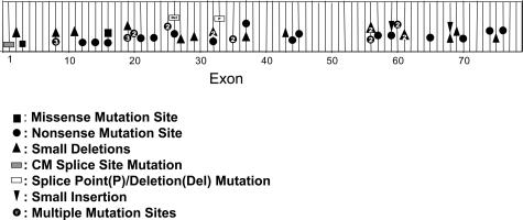
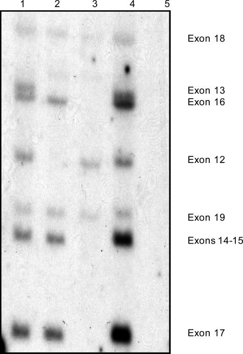
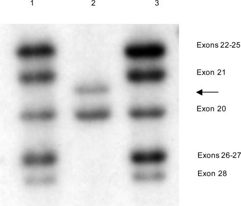
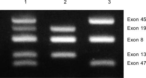
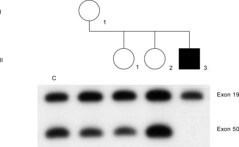
Mutations in the dystrophin gene result in both Duchenne and Becker muscular dystrophies (DMD and BMD). Approximately two-thirds of the affected patients have large deletions or duplications. Using the multiplex polymerase chain reaction and Southern blotting techniques, the detection of these larger mutations is relatively straightforward. Detection of the point mutations in the remaining one-third of the patients has been challenging, mainly due to the large gene size and lack of hotspots or prevalent mutations. However, with the addition of some of the newer molecular screening methods, it is becoming more feasible for clinical laboratories to test for point mutations in the larger genes like dystrophin. Here we review the clinical features, describe the mutation distributions, evaluate current molecular strategies, and illustrate how the genetic findings have impacted the current clinical diagnostics of Duchenne and Becker muscular dystrophies.


Figures

Figure 1
Figure 1
Distribution of deletions in the dystrophin gene in DMD and BMD patients. Each bar represents a deletion observed in a patient. The number to the right of the deletion bar indicates the number of independent patients sharing deletions of the same exon. Analysis was performed by Southern blotting and multiplex PCR. Arrows indicate the cDNA probes used for Southern blotting.
Figure 2
Figure 2
Distribution of duplications in the dystrophin gene in DMD and BMD patients. Each bar represents a duplication observed in a patient. The number right of the duplication bar indicates the number of independent patients sharing duplications of the same exon. Analysis was performed by Southern blotting. Arrows indicate the cDNA probes used for Southern blotting.
Figure 3
Figure 3
Distribution of point mutations in the dystrophin gene in DMD and BMD patients. The numbers indicate the number of independent patients sharing a mutation in the same exon. Table 1 specifies the actual nucleotide changes. CM, cardiomyopathy splice point mutation.
Figure 4
Figure 4
Southern hybridization using a dystrophin cDMD probe that hybridizes to exons 12 to 19 (DNA digested with HindIII). Lane 1 is an unaffected male control. The DMD patient in lane 2 has a gene deletion of exons 12 to 13. The DMD patient in lane 3 has a gene deletion of exons 13 to 17. The DMD patient in lane 4 has a gene duplication of exons 14 to 17. The DMD patient in lane 5 has a gene deletion of exons 12 to 19.
Figure 5
Figure 5
Southern hybridization using a dystrophin cDMD probe that hybridizes to exons 20 to 28 (DNA digested with HindIII). The DMD patient in lane 2 is deleted for exons 22 to 28 and an exon 21 junction fragment has resulted (arrow).
Figure 6
Figure 6
Multiplex DNA amplification of DNA from DMD patients. Lane 1, Unaffected male control. Lane 2, DMD patient deleted for exons 45 and 47. Lane 3, DMD patient deleted for exons 13 and 19.
Figure 7
Figure 7
Carrier determination by gene dosage. The affected son (II-3) is deleted for exon 50. Exon 19 is the internal standard because the affected son is not deleted for this exon. The mother (I-1) and the daughter (II-1) show a 50% reduction of hybridization of exon 50:19 dosage ratio compared to the noncarrier female control (c), and are therefore carriers. Daughter (II-2) is a noncarrier because her exon 50:19 dosage ratio is equivalent to the female control (c). The samples were subjected to 14 PCR cycles and the PCR products were Southern blotted and hybridized with the corresponding cDNA probes 2b-3 and 8.

Similar articles

Cited by

References

    1. Moser H. Duchenne muscular dystrophy: pathogenetic aspects and genetic prevention. Hum Genet. 1984;66:17–40. - PubMed
    1. Emery AEH. Muscle histology and creatine kinase levels in the fetus in Duchenne muscular dystrophy. Nature. 1977;266:472–473. - PubMed
    1. Emery AEH. New York: Oxford University Press; Duchenne Muscular Dystrophy. (ed 2.) 1993
    1. Farah MG, Evans EB, Vignos PJ. Echocardiographic evaluation of left function in Duchenne’s muscular dystrophy. Am J Med. 1980;69:248–252. - PubMed
    1. Leibowitz D, Dubowitz V. Intellect and behavior in Duchenne muscular dystrophy. Dev Med Neurol. 1981;23:577–590. - PubMed

Publication types