

Molecular cytogenetic analyses of immunoglobulin loci in nodular lymphocyte predominant Hodgkin's lymphoma reveal a recurrent IGH-BCL6 juxtaposition
- PMID: 16049307
- PMCID: PMC1867541
- DOI: 10.1016/S1525-1578(10)60564-8
Molecular cytogenetic analyses of immunoglobulin loci in nodular lymphocyte predominant Hodgkin's lymphoma reveal a recurrent IGH-BCL6 juxtaposition
Abstract
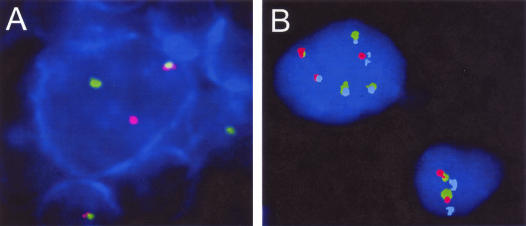
Chromosomal translocations juxtaposing different oncogenes to the immunoglobulin (IG) loci are the hallmark of various B-cell lymphomas. Because the tumor cells in nodular lymphocyte predominant Hodgkin's lymphoma (NLPHL) are also derived from B cells, we examined whether NLPHL harbors chromosomal translocations that affect IG loci. Fluorescence in situ hybridization was applied to 24 NLPHL cases using probes flanking the IGH, IGK, and IGL loci as well as the BCL6 gene. Fourteen of these cases were additionally analyzed by combined immunofluorescence and fluorescence in situ hybridization. Chromosomal breakpoints in the IGH locus were detected in five NLPHL. All these cases also contained a BCL6 breakpoint. Triple-color interphase cytogenetics demonstrated the presence of an IGH-BCL6 juxtaposition, indicating a t(3;14)(q27;q32) in all five cases. There was no evidence for breakpoints affecting the IGK or IGL loci. Our results show that translocations juxtaposing the BCL6 oncogene next to the IGH locus are recurrent in NLPHL.
Figures
Similar articles
-
Heterogeneity of BCL6 rearrangements in nodular lymphocyte predominant Hodgkin's lymphoma.Haematologica. 2004 Aug;89(8):965-72. Haematologica. 2004. PMID: 15339680
-
Interphase detection of BCL6/IgH fusion gene in non-Hodgkin lymphoma by fluorescence in situ hybridization.Cancer Genet Cytogenet. 1997 Dec;99(2):102-7. doi: 10.1016/s0165-4608(97)00203-3. Cancer Genet Cytogenet. 1997. PMID: 9398863
-
Molecular cytogenetic analysis of chromosomal breakpoints in the IGH, MYC, BCL6, and MALT1 gene loci in primary cutaneous B-cell lymphomas.J Invest Dermatol. 2004 Jul;123(1):213-9. doi: 10.1111/j.0022-202X.2004.22720.x. J Invest Dermatol. 2004. PMID: 15191563
-
Targeted somatic mutation of the BCL6 proto-oncogene and its impact on lymphomagenesis.Hematology. 2005 Apr;10(2):115-29. doi: 10.1080/10245330400026105. Hematology. 2005. PMID: 16019457 Review.
-
Splenic small B-cell lymphoma with IGH/BCL3 translocation.Hum Pathol. 2006 Feb;37(2):218-30. doi: 10.1016/j.humpath.2005.09.025. Hum Pathol. 2006. PMID: 16426923 Review.
Cited by
-
The RON receptor tyrosine kinase is a potential therapeutic target in Burkitt lymphoma.Cancer Biol Ther. 2013 Apr;14(4):370-7. doi: 10.4161/cbt.23718. Epub 2013 Jan 29. Cancer Biol Ther. 2013. PMID: 23360784 Free PMC article.
-
Is Hodgkin lymphoma just another B-cell lymphoma?Curr Hematol Malig Rep. 2009 Jul;4(3):125-8. doi: 10.1007/s11899-009-0018-1. Curr Hematol Malig Rep. 2009. PMID: 20425425 Review.
-
Coexisting and clonally identical classic hodgkin lymphoma and nodular lymphocyte predominant hodgkin lymphoma.Am J Surg Pathol. 2011 May;35(5):767-72. doi: 10.1097/PAS.0b013e3182147f91. Am J Surg Pathol. 2011. PMID: 21490448 Free PMC article.
-
Histopathologic Features and Differential Diagnosis in Challenging Cases of Nodular Lymphocyte Predominant B-cell Lymphoma/Nodular Lymphocyte Predominant Hodgkin Lymphoma.J Clin Transl Pathol. 2024 Jun;4(2):61-69. doi: 10.14218/jctp.2024.00015. J Clin Transl Pathol. 2024. PMID: 39070246 Free PMC article.
-
Genetic lesions in nodular lymphocyte-predominant Hodgkin lymphoma and T cell/histiocyte-rich large B-cell lymphoma identified by whole genome sequencing.Leukemia. 2025 Sep;39(9):2215-2225. doi: 10.1038/s41375-025-02679-3. Epub 2025 Jul 16. Leukemia. 2025. PMID: 40670673 Free PMC article.
References
-
- Hansmann ML, Stein H, Harris NL, Jaffe ES. Philadelphia: Lippincott Williams & Wilkins,; Pathology of Lymphocyte Predominant Hodgkin’s Disease. 1999
-
- Mason DY, Banks PM, Chan J, Cleary ML, Delsol G, de Wolf Peeters C, Falini B, Gatter K, Grogan TM, Harris NL. Nodular lymphocyte predominance Hodgkin’s disease. A distinct clinicopathological entity. Am J Surg Pathol. 1994;18:526–530. - PubMed
-
- Harris NL, Jaffe ES, Stein H, Banks PM, Chan JK, Cleary ML, Delsol G, De Wolf-Peeters C, Falini B, Gatter KC. A revised European-American classification of lymphoid neoplasms: a proposal from the International Lymphoma Study Group. Blood. 1994;84:1361–1392. - PubMed
-
- Küppers R, Rajewsky K, Bräuninger A, Hansmann ML. L&H cells in lymphocyte-predominant Hodgkin’s disease. N Engl J Med. 1998;338:763–765. - PubMed
Publication types
MeSH terms
Substances
LinkOut - more resources
Full Text Sources
Other Literature Sources
Medical
