

Autologous apoptotic cell engulfment stimulates chemokine secretion by vascular smooth muscle cells
- PMID: 16049322
- PMCID: PMC1603551
- DOI: 10.1016/S0002-9440(10)62980-X
Autologous apoptotic cell engulfment stimulates chemokine secretion by vascular smooth muscle cells
Abstract
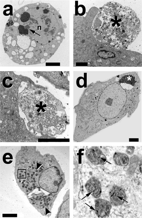
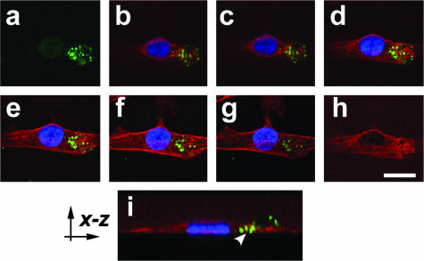
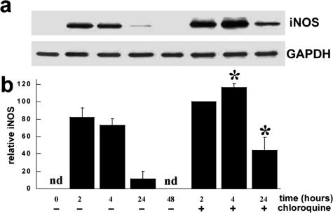
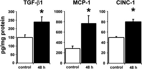
Apoptosis of vascular smooth muscle cells (VSMCs) occurs in vivo under both physiological and pathological settings. The clearance of apoptotic cells may be accomplished in part by the surrounding normal VSMCs. However, the fate of internalized apoptotic cells, the rate of intracellular degradation, and the consequences of these processes to VSMC biology are unknown. Electron microscopy and confocal fluorescence imaging showed that rat VSMCs effectively bound and internalized autologous apoptotic VSMCs in vitro. Within 2 hours, the internalized apoptotic cells were delivered to lysosomes, and the majority of these internalized cells and their proteins were efficiently degraded by 24 hours. After degradation was completed, the phagocytic VSMCs remained viable with normal rates of proliferation. Clearance of apoptotic cells by VSMCs did not induce the release of vascular wall matrix proteases but was associated with a 1.6-fold increase in transforming growth factor-beta1 release. Interestingly, clearance of apoptotic cells stimulated VSMCs to secrete monocyte-chemoattractant protein-1 and cytokine-induced neutrophil chemoattractant. The coordinated release of transforming growth factor-beta1 and chemokines suggests that autologous apoptotic cell clearance stimulates VSMCs to release molecules that specifically recruit professional phagocytes while simultaneously dampening the inflammatory response and preventing vascular injury.
Figures
Similar articles
-
Vascular smooth muscle cell apoptosis induces interleukin-1-directed inflammation: effects of hyperlipidemia-mediated inhibition of phagocytosis.Circ Res. 2010 Feb 5;106(2):363-72. doi: 10.1161/CIRCRESAHA.109.208389. Epub 2009 Nov 19. Circ Res. 2010. PMID: 19926874
-
JE mRNA accumulates rapidly in aortic injury and in platelet-derived growth factor-stimulated vascular smooth muscle cells.Circ Res. 1992 Feb;70(2):314-25. doi: 10.1161/01.res.70.2.314. Circ Res. 1992. PMID: 1735132
-
Downregulation of nitric oxide accumulation by cyclooxygenase-2 induction and thromboxane A2 production in interleukin-1beta-stimulated rat aortic smooth muscle cells.J Hypertens. 2002 Mar;20(3):455-61. doi: 10.1097/00004872-200203000-00021. J Hypertens. 2002. PMID: 11875313
-
Urotensin II-induced collagen synthesis in cultured smooth muscle cells from rat aortic media and a possible involvement of transforming growth factor-β1/Smad2/3 signaling pathway.Regul Pept. 2013 Mar 10;182:53-8. doi: 10.1016/j.regpep.2012.12.006. Epub 2013 Feb 9. Regul Pept. 2013. PMID: 23403244
-
Vascular smooth muscle cell death, autophagy and senescence in atherosclerosis.Cardiovasc Res. 2018 Mar 15;114(4):622-634. doi: 10.1093/cvr/cvy007. Cardiovasc Res. 2018. PMID: 29360955 Review.
Cited by
-
Clearance of Dying Cells by Phagocytes: Mechanisms and Implications for Disease Pathogenesis.Adv Exp Med Biol. 2016;930:25-49. doi: 10.1007/978-3-319-39406-0_2. Adv Exp Med Biol. 2016. PMID: 27558816 Free PMC article. Review.
-
Efferocytosis of vascular cells in cardiovascular disease.Pharmacol Ther. 2022 Jan;229:107919. doi: 10.1016/j.pharmthera.2021.107919. Epub 2021 Jun 23. Pharmacol Ther. 2022. PMID: 34171333 Free PMC article. Review.
-
IFN-gamma primes intact human coronary arteries and cultured coronary smooth muscle cells to double-stranded RNA- and self-RNA-induced inflammatory responses by upregulating TLR3 and melanoma differentiation-associated gene 5.J Immunol. 2010 Jul 15;185(2):1283-94. doi: 10.4049/jimmunol.0902283. Epub 2010 Jun 18. J Immunol. 2010. PMID: 20562257 Free PMC article.
-
Trophoblast-mediated spiral artery remodelling: a role for apoptosis.J Anat. 2009 Jul;215(1):21-6. doi: 10.1111/j.1469-7580.2008.01039.x. Epub 2009 Feb 9. J Anat. 2009. PMID: 19215319 Free PMC article. Review.
-
Macrophages in Atherosclerosis, First or Second Row Players?Biomedicines. 2021 Sep 13;9(9):1214. doi: 10.3390/biomedicines9091214. Biomedicines. 2021. PMID: 34572399 Free PMC article. Review.
References
-
- Devlin AM, Clark JS, Reid JL, Dominiczak AF. DNA synthesis and apoptosis in smooth muscle cells from a model of genetic hypertension. Hypertension. 2000;36:110–115. - PubMed
-
- Bennett MR, Evan GI, Schwartz SM. Apoptosis of rat vascular smooth muscle cells is regulated by p53-dependent and -independent pathways. Circ Res. 1995;77:266–273. - PubMed
Publication types
MeSH terms
Substances
Grants and funding
LinkOut - more resources
Full Text Sources
Medical
Research Materials
