. 2005 Aug;167(2):355-63.
doi: 10.1016/S0002-9440(10)62981-1.

Glomerular aging in females is a multi-stage reversible process mediated by phenotypic changes in progenitors

Affiliations

Glomerular aging in females is a multi-stage reversible process mediated by phenotypic changes in progenitors

Zheng Feng et al. Am J Pathol. 2005 Aug.

Abstract

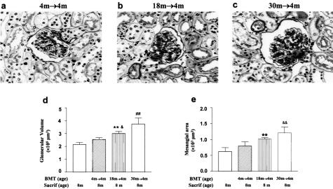
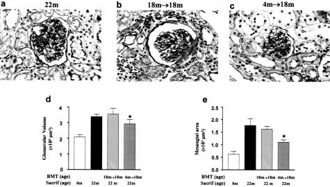
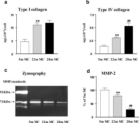
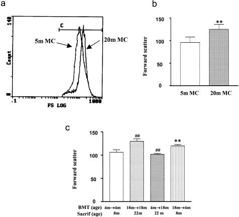
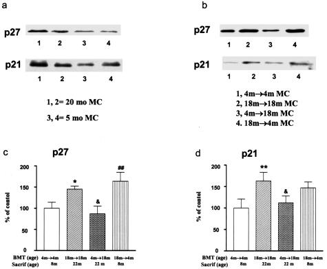
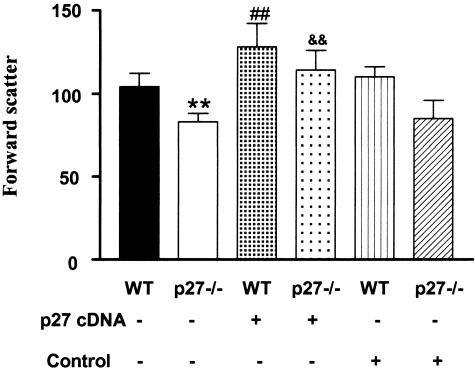
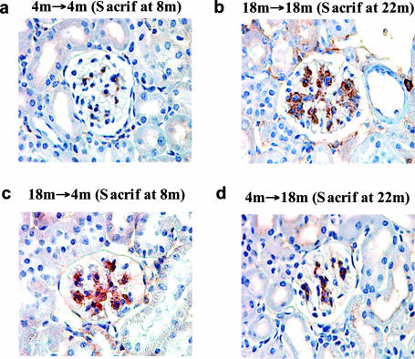
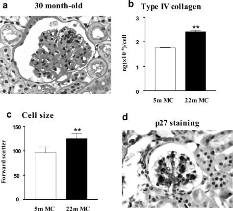
The glomeruli of postmenopausal C57BL6 mice, and age-matched males, show progressive hypertrophy and glomerulosclerosis. We asked whether this was a multistage process, was due to alterations in glomerular progenitors, and was reversible in female mice. Using cross bone marrow transplants (BMT) between young and old females, we found that BMT delivered a phenotype that was donor age-specific. The fact that lesions in young recipients were more severe if the donors were in late rather than early menopause suggested that new progenitor phenotypes had appeared. Postmenopausal recipients of BMT from young donors had reduced glomerular hypertrophy and sclerosis, implying that the aging lesions in females were reversible and that progenitors, rather than the local environment, determined the glomerular profile. The altered phenotype included increased extracellular matrix synthesis and decreased matrix metalloproteinase-2 levels as well as cell hypertrophy. The mechanism of the cellular hypertrophy was due to uncoupling of hypertrophy from proliferation, resulting from elevated p27 levels. Thus, glomerular hypertrophy and sclerosis in aging females is a multistage process, is reversible, and may be determined by the phenotype of bone marrow-derived progenitor cells.


Figures

Figure 1
Figure 1
Transfer of the aging lesions after BMT. a: The glomeruli of 4-month-old recipients of 4-month-old BMT (4 months → 4 months, age at BMT = 4 months; age at sacrifice = 8 months) were normal by light microscopy. Sacrif, sacrifice; m, month. PAS stain. b: The mesangial areas, especially at the vascular poles, were enlarged in 4-month-old recipients of 18-month-old BMT (18 months → 4 months). c: Diffuse expansion of the mesangial areas was present in 4-month-old recipients of 30-month-old BMT (30 months → 4 months). d: The glomerular volume was similar in 4 months → 4 months and 8-month-old naïve controls. The glomerular volume in 18 months → 4 months was larger than that in 8 months (**P < 0.01) and in 4 months → 4 months (&P < 0.05). The glomerular volume in 30 months → 4 months, compared with either young controls (8 months and 4 months → 4 months) (##P < 0.01) or with 18 months → 4 months, was further increased (##P < 0.01). e: The mesangial areas in 4 months → 4 months and 8 months were similar. However, mesangial area was increased in 18 months → 4 months compared with 8 months (**P < 0.01), and was further increased in 30 months → 4 months as compared with 4 months → 4 months (&&P < 0.01). The mesangial area did not differ between 4 months → 4 months and 18 months → 4 months. Original magnification, ×400 (a–c).
Figure 2
Figure 2
Modulation of glomerular structure after BMT. a: Glomerular hypertrophy and expansion of the extracellular matrix, tubular atrophy, thickened tubular basement membranes, and interstitial inflammatory cell infiltration were present in 22-month-old naïve mice (22 months). PAS stain. b: Glomerular and tubulointerstitial lesions were prominent in 18-month-old recipients of BMT from 18-month-old mice (18 months → 18 months, age at BMT = 18 months; age at sacrifice = 22 months), changes similar to that of 22-month-old naïve mice. Sacrif, sacrifice; m, month. c: Glomerular hypertrophy and extracellular matrix expansion were less evident, and tubulointerstitial lesions were less severe, in 18-month-old recipients of BMT from 4-month-old mice (4 months → 18 months). d: Glomerular hypertrophy was significantly decreased in 4 months → 18 months compared to 22 months or 18 months → 18 months (*P < 0.05). e: Mesangial area was decreased in 4 months → 18 months compared with the 22 months or 18 months → 18 months (*P < 0.05). The mesangial areas of 22 months and 18 months →18 months were similar. Original magnification, ×400 (a–c).
Figure 3
Figure 3
Progressive phenotypic changes in ECM synthesis and degradation in mesangial cells from aging females. a: Mesangial cells isolated from 22- and 28-month-old mice (22-month MC, 28-month MC) produced significantly more type I collagen than mesangial cells isolated from 5-month-old mice (5-month MC). **P < 0.01. b: Progressive increase in type IV collagen production in mesangial cells with aging (**P < 0.01 versus 5-month MC; ##P < 0.01 versus 22-month MC). c: MMP-2 activity was reduced in mesangial cells from 22-month-old mice (22-month MC) and it was further decreased in mesangial cells from 28-month-old mice. d: Quantitation of three independent experiments reveals that the production of MMP-2 activity by mesangial cells progressively decreased with aging (**P < 0.01 versus 5-month MC; ##P < 0.01 versus 22-month MC).
Figure 4
Figure 4
Analysis of mesangial cell size by flow cytometry. a: Forward scatter, an index of cell size, revealed that mesangial cells from 20-month-old mice (20-month MC) were larger than mesangial cells from 5-month-old mice (5-month MC). b: Analyses of three independent measurements of two cell lines from 5-month-old mice and two cell lines from 20-month-old mice confirm the increase in cell size. **P < 0.01. c: Mesangial cells from 18-month-old recipients of BMT from 18-month-old mice donors (18 months → 18 months, age at BMT = 18 months; age at sacrifice = 22 months) were larger than those from 4-month-old recipients of BMT from 4-month-old donors (4 months → 4 months, P < 0.01). Mesangial cells from 4-month-old recipients of BMT from 18-month-old donors (18 months → 4 months) were larger than 4-month-old recipients of BMT from 4-month-old donors (**P < 0.01). On the other hand, mesangial cells from 18-month-old recipients of BMT from 4-month-old donors (4 months → 18 months) were reduced in size compared to 18-month-old recipients of BMT from 18-month-old donors (##P < 0.01).
Figure 5
Figure 5
p27 and p21 and mesangial cell size in aging. a: A representative Western blot of two cell lines demonstrating higher p27 levels in mesangial cells isolated from 20-month-old mice (20-month MC) (lanes 1 and 2) compared to mesangial cells isolated from 5-month-old mice (5-month MC) (lanes 3 and 4). There is also a slight increase in p21 levels. b: Representative Western blot gels demonstrating that p27 levels were increased in mesangial cells from 4-month-old recipients of BMT from 18-month-old donors (18 month → 4-month MC, age at BMT = 4 months; age at sacrifice = 8 months). However, p27 levels were decreased in mesangial cells isolated from 18-month-old recipients of BMT from 4-month-old donors (4 month → 18 month MC), compared to their respective age-matched transplant controls (18 month → 18 month MC or 4 month → 4 month MC). Note that p21 levels follow p27 levels, but are less pronounced. c: Densitometry scanning of Western blot gels from three experiments revealed that p27 levels were increased in mesangial cells from 18-month-old recipients of BMT from 18-month-old donors (18 month → 18 month MC) and were decreased in 18-month-old recipients of BMT from 4-month-old donors (4 month → 18 month MC). Data obtained from 4 month → 4 month MC were defined as 100% (control). *P < 0.05 versus 4 month → 4 month MC; &P < 0.05 versus 18 month → 18 month MC; ##P < 0.01 versus 4 month → 4 month MC. d: Densitometry scanning of Western blot gels from three experiments revealed that p21 levels were increased in mesangial cells from 18-month-old recipients of BMT from 18-month-old donors and were decreased in 18-month-old recipients of BMT from 4-month-old donors. Data obtained from 4 month → 4 month MC were defined as 100% (control). **P < 0.01 versus 4 month → 4 month MC; &P < 0.05 versus 18 month → 18 month MC.
Figure 6
Figure 6
Relationship between p27 and mesangial cell size, general aspects. Cell size analyzed by flow cytometry revealed that mesangial cells isolated from p27 knockout mice were smaller than those isolated from wild-type mice (WT, **P < 0.01). Transfection of p27−/− mesangial cells with a p27 cDNA expression vector restored cells to a size comparable with that of wild-type (WT) mesangial cells (&&P < 0.01 versus p27−/−). Wild-type mesangial cells transfected with the p27 cDNA expression vector were also increased in size (##P < 0.01 versus WT or WT transfected with an empty vector). No change in cell size was found in mesangial cells transfected with an empty vector.
Figure 7
Figure 7
In vivo correlations between p27 immunostaining and glomerular size. a: There is a small, scattered amount of p27 (brown staining material) in this glomerulus from a 4-month-old recipient of a BMT from 4-month-old donors (4 months → 4 months). b: A glomerulus from an 18-month-old recipient of a BMT from 18-month-old donors (18 months → 18 months) contains a relatively large amount of stainable p27. c: A glomerulus from a 4-month-old recipient of a BMT from 18-month-old donors (18 months → 4 months) shows a similar amount of stainable p27 to that in b. d: A glomerulus from an 18-month-old recipient of BMT from 4-month-old donors (4 months → 18 months) has less stainable p27 than in either b or c.
Figure 8
Figure 8
Glomerular lesions and phenotypic changes in mesangial cells from aging males. a: Glomerular size, mesangial area, and cell number were increased in 30-month-old male B6 mouse (PAS). b: Mesangial cells isolated from 22-month-old male B6 mouse produced significantly more type IV collagen than mesangial cells isolated from 5-month-old male B6 mouse (**P < 0.01). c: Mesangial cells isolated from 22-month-old male B6 mouse were larger than mesangial cells isolated from 5-month-old male B6 mouse (**P < 0.01). d: A glomerulus from a 22-month-old male B6 mouse shows a large amount of p27 staining. Original magnification ×400 (a).

Similar articles

Cited by

References

    1. Coresh J, Astor BC, Greene T, Eknoyan G, Levey AS. Prevalence of chronic kidney disease and decreased kidney function in the adult US population: Third National Health and Nutrition Examination Survey. Am J Kidney Dis. 2003;41:1–12. - PubMed
    1. US Renal Data System Washington, DC: The National Institutes of Health, NIDDK; USRDS 1999 Annual Report. 1999
    1. Zheng F, Plati AR, Potier M, Schulman Y, Berho M, Banerjee A, Leclercq B, Zisman A, Striker LJ, Striker GE. Resistance to glomerulosclerosis in B6 mice disappears after menopause. Am J Pathol. 2003;162:1339–1348. - PMC - PubMed
    1. Cornacchia F, Fornoni A, Plati AR, Thomas A, Wang Y, Inverardi L, Striker LJ, Striker GE. Glomerulosclerosis is transmitted by bone marrow-derived mesangial cell progenitors. J Clin Invest. 2001;108:1649–1656. - PMC - PubMed
    1. Zheng F, Cornaccia F, Schulman I, Banerjee A, Cheng QL, Potier M, Plati AR, Berho M, Elliot SJ, Li J, Fornoni A, Zang YJ, Zisman A, Striker LJ, Striker GE. Development of albuminuria and glomerular lesions in normoglycemic B6 recipients of db/db mice bone marrow: the role of mesangial cell progenitors. Diabetes. 2004;53:2420–2427. - PubMed

Publication types

MeSH terms

Substances