. 2005 Aug;167(2):365-79.
doi: 10.1016/S0002-9440(10)62982-3.

Loss of fibroblast Thy-1 expression correlates with lung fibrogenesis

Affiliations

Loss of fibroblast Thy-1 expression correlates with lung fibrogenesis

James S Hagood et al. Am J Pathol. 2005 Aug.

Abstract

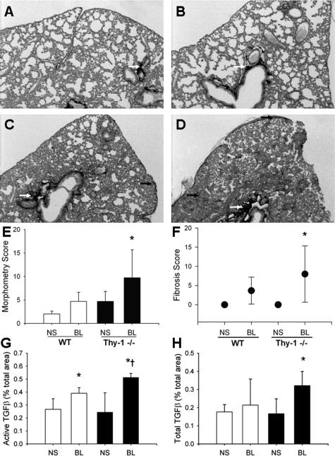
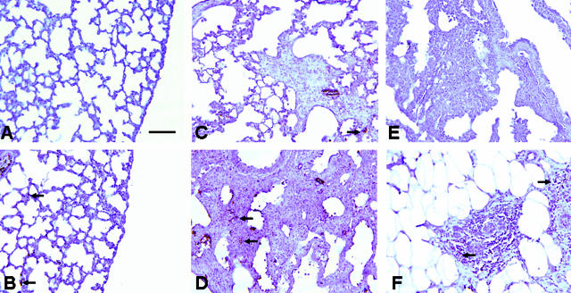
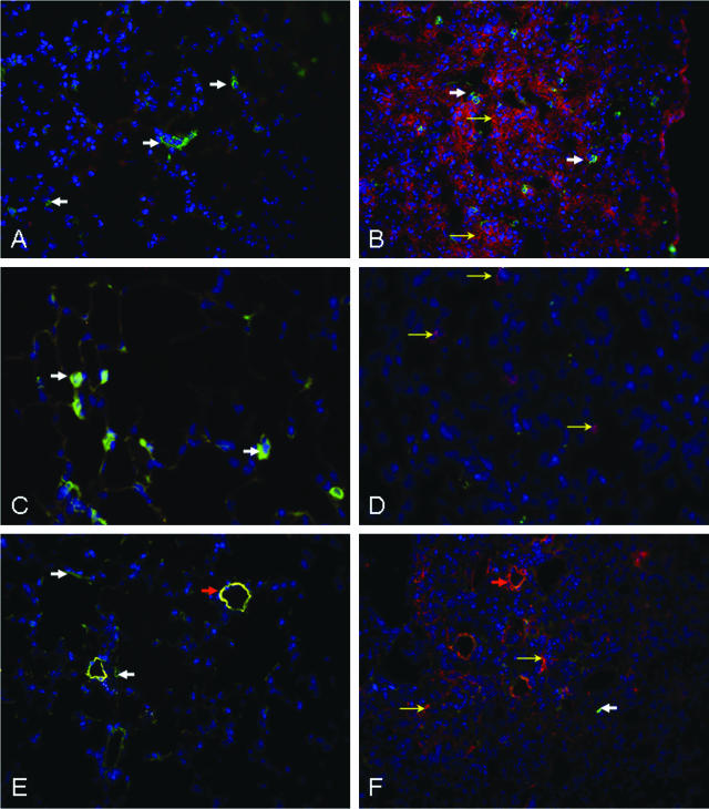
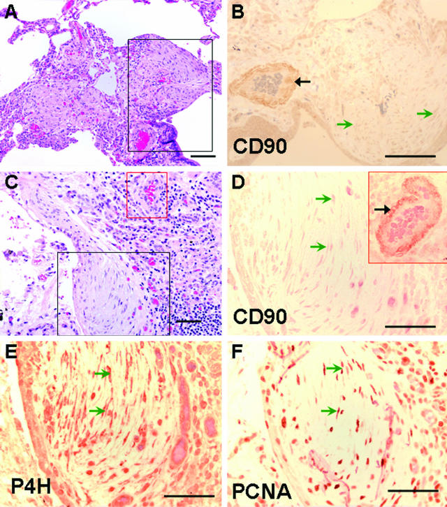
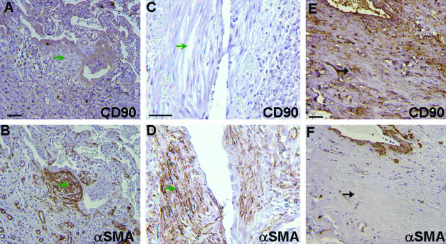
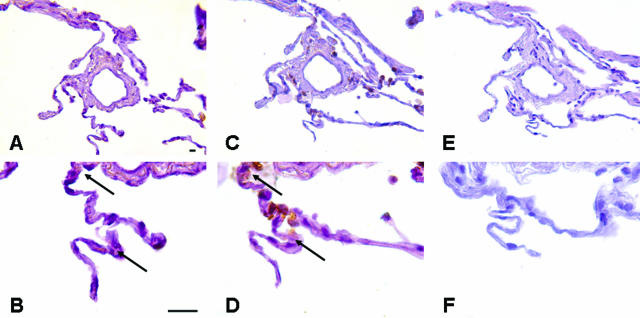
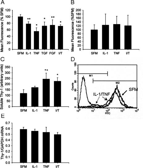
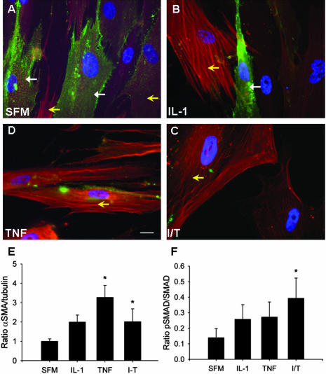
Fibroblasts consist of heterogeneous subpopulations that have distinct roles in fibrotic responses. Previously we reported enhanced proliferation in response to fibrogenic growth factors and selective activation of latent transforming growth factor (TGF)-beta in fibroblasts lacking cell surface expression of Thy-1 glycoprotein, suggesting that Thy-1 modulates the fibrogenic potential of fibroblasts. Here we report that compared to controls Thy-1-/- C57BL/6 mice displayed more severe histopathological lung fibrosis, greater accumulation of lung collagen, and increased TGF-beta activation in the lungs 14 days after intratracheal bleomycin. The majority of cells demonstrating TGF-beta activation and myofibroblast differentiation in bleomycin-induced lesions were Thy-1-negative. Histological sections from patients with idiopathic pulmonary fibrosis demonstrated absent Thy-1 staining within fibroblastic foci. Normal lung fibroblasts, in both mice and humans, were predominantly Thy-1-positive. The fibrogenic cytokines interleukin-1 and tumor necrosis factor-alpha induced loss of fibroblast Thy-1 surface expression in vitro, which was associated with Thy-1 shedding, Smad phosphorylation, and myofibroblast differentiation. These results suggest that fibrogenic injury promotes loss of lung fibroblast Thy-1 expression, resulting in enhanced fibrogenesis.


Figures

Figure 1
Figure 1
Thy-1−/− mice have increased fibrosis and lung collagen accumulation after intratracheal BL. A–D: Representative picrosirius red-stained 5-μm sections of formalin-fixed, paraffin-embedded right lungs of 6-week-old C57BL/6 WT (A, C) or Thy-1−/− (B, D) mice that received intratracheal BL (4 U/kg; C and D) or an equal volume of normal saline (A, B) 14 days previously, as described in Materials and Methods. White arrows indicate picrosirius red-stained collagen in peribronchial areas, black arrows indicate collagen in subpleural and interstitial areas. E–H: Picrosirius red-stained lungs were analyzed under polarized light for morphometric analysis of collagen in a blinded manner as described in Materials and Methods. Mean ± SD are shown [E: *P < 0.01 for Thy-1−/− BL (n = 8) versus WT NS (n = 5) and Thy-1−/− NS (n = 8), P < 0.05 versus WT BL (n = 8)]. Trichrome-stained lungs from the same experiment were examined by an experienced veterinary pathologist (T.R.S.) in a blinded manner and assigned a histopathological score for fibrosis as described in Materials and Methods (F: *P < 0.05 versus NS-treated controls). IHC staining for active and total TGF-β was performed and analyzed using morphometric analysis as described in Materials and Methods (G: *P < 0.01 versus NS-treated controls, P = 0.012 versus WT BL; H: *P < 0.005 versus all other conditions). Original magnifications, ×2.
Figure 2
Figure 2
Lungs from Thy-1−/− mice demonstrate increased immunoreactivity for phosphorylated Smad 2/3, indicative of TGF-β activity, after intratracheal BL. Photomicrographs of representative (5 μm) sections from formalin-fixed, paraffin-embedded right lungs of 6-week-old C57BL/6 WT (A, C) or Thy-1−/− (B, D) mice that received intratracheal BL (C, D; 4 U/kg) or an equal volume of saline (A, B) 14 days previously, stained with antibody to phosphorylated Smad 2/3 (pSmad2/3), as described in Materials and Methods. Arrows indicate areas of strong positivity (dark brown staining). E: Section from BL-treated KO mouse from the same experiment, stained with control antiserum; F: section from invasive breast carcinoma stained with pSmad2/3 antibody. Scale bar, 100 μm. Original magnifications, ×20.
Figure 3
Figure 3
Lungs from WT mice demonstrate immunoreactivity for active TGF-β, BrdU, and α-SMA, in cells lacking Thy-1 expression, in fibrotic areas after intratracheal BL. Immunofluorescence photomicrographs of representative (4 μm) cryostat sections from frozen, OCT-embedded lungs of 6-week-old C57BL/6 WT mice that received intratracheal BL (B, D, F; 4 U/kg) or an equal volume of saline (A, C, E), 14 days previously, and that received 1 mg of BrdU intraperitoneally 90 minutes before sacrifice. All sections were stained with FITC-coupled antibody to Thy-1 (CD90.2, green) and Hoechst 33258 (blue). A, B: Sections also stained with active TGF-β antibody. Solid white arrows indicate Thy-1-positive cells (green), open yellow arrows indicate cells positive for active TGF-β (red). C, D: Sections also stained with BrdU antibody. Solid white arrows indicate Thy-1-positive cells (green), open yellow arrows indicate BrdU-positive cells (magenta). E, F: Sections also stained with α-SMA antibody. Solid white arrows in E and F indicate Thy-1-positive cells (E, green), open yellow arrows indicate α-SMA-positive fibroblasts (F, red). Orange arrows (E, F) indicate small blood vessels in which Thy-1 and α-SMA co-localize in saline-treated animals (E), but only α-SMA is seen in BL-treated animals (F). Original magnifications: ×25 (A, B, E, F); ×40 (C, D).
Figure 4
Figure 4
Fibroblasts within fibroblastic foci demonstrate absent Thy-1 (CD90) staining. Photomicrographs of serial sections from lung biopsy of two patients with IPF/UIP. A:: H&E. Black box demarcates a fibroblastic focus and correlates to area shown in B. B: Thy-1 (CD90). Positive immunostaining is seen predominantly in the wall of a vascular structure (black arrow). Spindle-shaped cells in fibroblastic foci (open green arrowheads) demonstrate absent staining for Thy-1 and are equivalent to isotype control (not shown). C: H&E. Section from another IPF patient. Black box demarcates a fibroblastic focus and correlates to area shown in D–F. Red box correlates to inset in D. D: Thy-1 (CD90). Positive immunostaining is seen predominantly in capillaries and in the wall of a vascular structure (artery, red box) within the same section (solid black arrow). E: IHC for P4H. Dark reddish brown staining in spindle-shaped cells indicates fibroblasts (open green arrowheads). F: IHC for PCNA. Dark reddish brown nuclei indicate proliferating cells (open green arrowheads). Scale bars, 132.8 μm. Original magnifications: ×10 (A, C); ×20 (B, D–F).
Figure 5
Figure 5
Myofibroblasts within fibroblastic foci demonstrate absent Thy-1 staining. Photomicrographs of sections from lung biopsies of three patients with UIP/IPF. Top: IHC for Thy-1 (CD90). Bottom: IHC for α-SMA from nearby sections from the same patients. A–D: Fibroblastic foci showing absent Thy-1/CD90 staining in myofibroblastic cells (open green arrowheads). E, F: Area of dense fibrosis showing Thy-1/CD90-positive spindle-shaped cells that are negative for α-SMA (solid black arrows). Scale bar, 50 μm. Original magnifications, ×25 (A, B, E, F); ×40 (C, D).
Figure 6
Figure 6
Normal lungs demonstrate Thy-1 staining in a pattern consistent with normal fibroblasts. A–F: Photomicrographs of serial sections from normal lung. A, B: IHC for Thy-1 (CD90). Dark brown staining is seen in cytoplasm and nuclei of spindle-shaped cells in the adventitia of a small blood vessel and in interstitium of alveolar septae (arrows). C, D: IHC for P4H. Dark brown staining is seen in a pattern similar to that observed for CD90. E, F: Control antiserum does not stain normal lung cells. Scale bar, 20 μm. Original magnifications, ×20 (A, C, E); ×100 (B, D, F).
Figure 7
Figure 7
Fibrogenic cytokines induce loss of cell surface Thy-1 staining in lung fibroblasts and accumulation of soluble Thy-1 in conditioned medium. A: Normal lung fibroblasts were cultured in the presence of IL-1β (5 ng/ml), TNF-α (1 ng/ml), combined IL-1β and TNF-α (I/T, 5 ng/ml and 1 ng/ml, respectively), FGF-1 (20 ng/ml), or TGF-β (1 ng/ml) for 96 hours, with fresh cytokine-containing medium or SFM replaced daily. Cells were resuspended, stained with anti-CD90 FITC, and analyzed by flow cytometry. The mean FITC fluorescence of the positive population (eg, M2 in D) was averaged for n = 3 per condition and expressed relative to values for SFM-exposed cells, arbitrarily set at 100%. *P < 0.001 versus SFM; +P < 0.001 versus TNF; < 0.001 versus TNF and P = 0.007 versus IL-1. B: After 96 hours of cytokine exposure, monolayers were washed three times with SFM and cultured for an additional 72 hours in SFM alone, then resuspended and analyzed by flow cytometry. The mean FITC fluorescence for the positive population was averaged and expressed relative to values for SFM-exposed cells, arbitrarily set at 100%. C: Thy-1 was immunoprecipitated from conditioned media of fibroblasts exposed to IL-1β (5 ng/ml), TNF-α (1 ng/ml), or combined IL-1β and TNF-α (T+I, 5 ng/ml and 1 ng/ml, respectively) for 72 hours, as above. Band density of enhanced chemiluminescent images was averaged for three separate experiments. Bars represent mean ± SD. *P < 0.05 versus SFM; +P < 0.05 versus IL-1. D: Normal fibroblasts were cultured in the presence of IL-1 and TNF (5 and 1 ng/ml, respectively) for 72 hours. CD90 expression was analyzed by flow cytometry. A representative histogram indicated by solid arrow indicates CD90 staining of SFM-exposed cells, the histogram indicated by broken arrows indicates IL-1/TNF-exposed cells, the histogram at the far left indicates SFM-exposed cells stained with isotype control Ab. Dashed arrow, open arrowhead indicates leftward shift of FITC staining intensity and dotted arrow, closed arrowhead indicates negatively staining cells. E: RNA was prepared from fibroblasts cultured in the presence of IL-1 and/or TNF for 6 hours and subjected to RT-PCR using primers specific for human Thy-1 and GAPDH as described in Materials and Methods. Histogram depicts the ratio of band intensity for Thy-1/GAPDH PCR products averaged for three experiments.
Figure 8
Figure 8
Loss of Thy-1 expression in human lung fibroblasts correlates with increased expression of α-SMA and pSmad 2/3. Normal lung fibroblasts were cultured in the presence of IL-1β and TNF-α, alone or in combination, as in Figure 7, above, for 72 hours. A–D: Immunofluorescence photomicrographs of fibroblasts grown on coverslips and stained with FITC-conjugated Thy-1 antibody and Cy3-conjugated α-SMA antibody. Closed arrows indicate cell-surface Thy-1 expression (green), open yellow arrows indicate intracellular α-SMA (red). E and F: Densitometric analysis of immunoblots for α-SMA (expressed as ratio to tubulin, E) and pSmad2/3 (expressed as a ratio to total Smad, F). *P < 0.05 versus SFM. Scale bar, 20 μm. Original magnifications, ×63.

Similar articles

Cited by

References

    1. Crystal RG, Bitterman PB, Mossman B, Schwarz MI, Sheppard D, Almasy L, Chapman HA, Friedman SL, King TE, Jr, Leinwand LA, Liotta L, Martin GR, Schwartz DA, Schultz GS, Wagner CR, Musson RA. Future research directions in idiopathic pulmonary fibrosis: summary of a National Heart, Lung, and Blood Institute working group. Am J Respir Crit Care Med. 2002;166:236–246. - PubMed
    1. King TE, Schwarz MI, Brown K, Tooze JA, Colby TV, Waldron JA, Flint A, Thurlbeck W, Cherniack RM. Idiopathic pulmonary fibrosis. Relationship between histopathologic features and mortality. Am J Respir Crit Care Med. 2001;164:1025–1032. - PubMed
    1. Nicholson AG, Fulford LG, Colby TV, du Bois RM, Hansell DM, Wells AU. The relationship between individual histologic features and disease progression in idiopathic pulmonary fibrosis. Am J Respir Crit Care Med. 2002;166:173–177. - PubMed
    1. Katzenstein AL, Myers JL. Idiopathic pulmonary fibrosis: clinical relevance of pathologic classification. Am J Respir Crit Care Med. 1998;157:1301–1315. - PubMed
    1. Selman M, King TE, Pardo A. Idiopathic pulmonary fibrosis: prevailing and evolving hypotheses about its pathogenesis and implications for therapy. Ann Intern Med. 2001;134:136–151. - PubMed

Publication types

MeSH terms