Comparative Study
. 2005 Aug;167(2):409-17.
doi: 10.1016/s0002-9440(10)62985-9.

Proliferation of estrogen receptor-alpha-positive mammary epithelial cells is restrained by transforming growth factor-beta1 in adult mice

Affiliations
Comparative Study

Proliferation of estrogen receptor-alpha-positive mammary epithelial cells is restrained by transforming growth factor-beta1 in adult mice

Kenneth B R Ewan et al. Am J Pathol. 2005 Aug.

Abstract

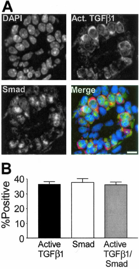
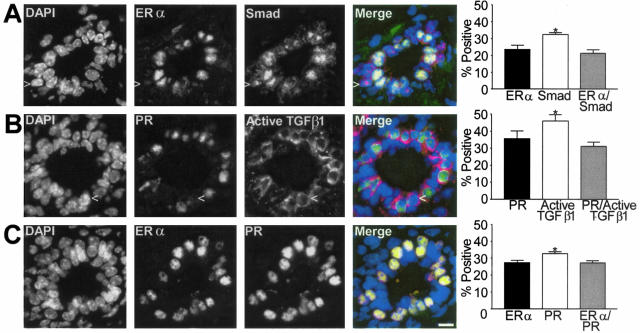
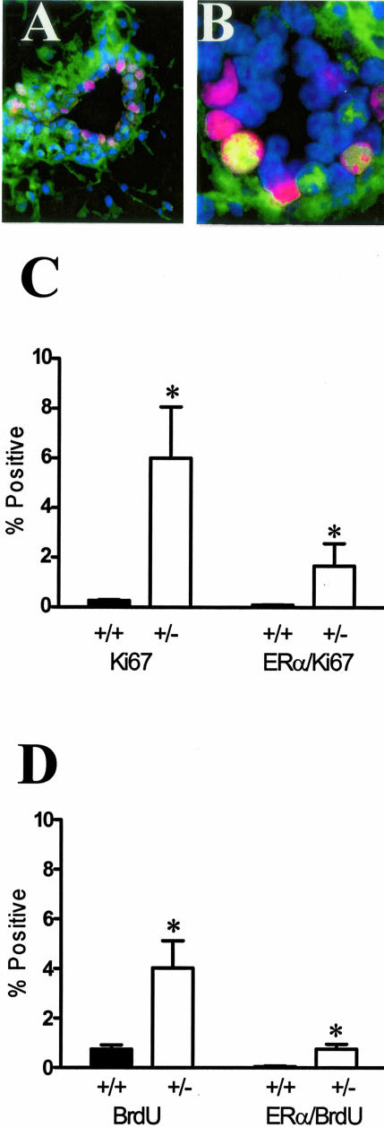
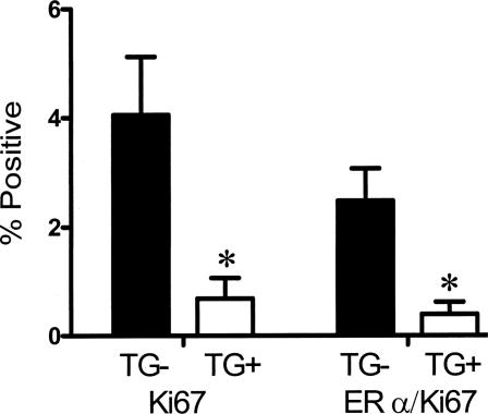
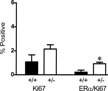
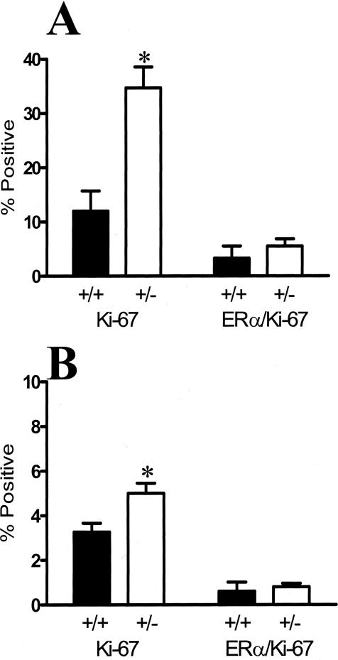
Transforming growth factor (TGF)-beta1 is a potent inhibitor of mammary epithelial proliferation. In human breast, estrogen receptor (ER)-alpha cells rarely co-localize with markers of proliferation, but their increased frequency correlates with breast cancer risk. To determine whether TGF-beta1 is necessary for the quiescence of ER-alpha-positive populations, we examined mouse mammary epithelial glands at estrus. Approximately 35% of epithelial cells showed TGF-beta1 activation, which co-localized with nuclear receptor-phosphorylated Smad 2/3, indicating that TGF-beta signaling is autocrine. Nuclear Smad co-localized with nuclear ER-alpha. To test whether TGF-beta inhibits proliferation, we examined genetically engineered mice with different levels of TGF-beta1. ER-alpha co-localization with markers of proliferation (ie, Ki-67 or bromodeoxyuridine) at estrus was significantly increased in the mammary glands of Tgf beta1 C57/bl/129SV heterozygote mice. This relationship was maintained after pregnancy but was absent at puberty. Conversely, mammary epithelial expression of constitutively active TGF-beta1 via the MMTV promoter suppressed proliferation of ER-alpha-positive cells. Thus, TGF-beta1 activation functionally restrains ER-alpha-positive cells from proliferating in adult mammary gland. Accordingly, we propose that TGF-beta1 dysregulation may promote proliferation of ER-alpha-positive cells associated with breast cancer risk in humans.


Figures

Figure 1
Figure 1
Nuclear R-Smad protein co-localizes in mammary epithelial cells that are positive for active TGF-β1. A: Individual channel images of DAPI-stained nuclei (DAPI), R-Smad, and TGF-β1 immunostaining in a transverse section of a duct. Merged image shows R-Smad immunoreactivity as green and active TGF-β1 as red. B: Active TGF-β1 and nuclear R-Smad are markers of the same cell population in the mammary epithelium at estrus. Shown are means ± SEM from three C57BL/6–129SV mixed background animals. At least 250 cells were scored per animal. Scale bar, 20 μm.
Figure 2
Figure 2
Subpopulations of the mammary epithelium of the C57BL/6–129SV mixed background mouse at estrus. A: Example of dual-immunofluorescence localization of ER-α and R-Smad immunoreactivity shows the gray scale individual images of DAPI-stained nuclei, ER-α, R-Smad, and a merged color image showing ER-α immunoreactivity as green and nuclear Smad as red which makes nuclei positive for both appear yellow/orange. Column graph shows mean co-localization frequency ± SEM (n = 3). B: Gray scale images of DAPI-stained nuclei, PR, and active TGF-β1 immunostaining in ductal epithelium. The merged color image shows that most PR-positive cells (green nuclei) are also positive for cytoplasmic active TGF-β1 (red). Column graph shows mean co-localization frequency ± SEM (n = 3). C: Individual channels and merged image of ER-α and PR immunoreactivity in a transverse section of a duct. ER-α-positive cells all exhibit PR and the nuclei appear orange in the merged image. Column graph shows mean co-localization frequency ± SEM (n = 3). Scale bar, 20 μm.
Figure 3
Figure 3
TGF-β1 depletion results in increased frequency of ER-α-positive mammary epithelial cells in cycle. A: Dual-immunofluorescence localization of ER-α (green; nonnuclear staining is nonspecific) and Ki67 (red) in mammary epithelium. Nuclei are counterstained with DAPI (blue). B: Dual-immunofluorescence localization of ER-α and Ki67, as in A, of higher magnification of a mammary duct with an example of double-labeled nucleus (yellow). C: Ki-67 and ER-α/Ki-67 co-localization frequency in mammary epithelium of nulliparous Tgfβ1 heterozygote (+/− in figure) and Tgfβ1 wild-type (+/+) C57BL/6–129SV mice at estrus. Three animals per genotype and at least 300 cells per animal were scored for presence of ER-α and Ki-67 immunoreactivity. Asterisks indicate significant difference from Tgfβ1 wild-type mean frequency (P < 0.01; t-test). D: BrdU and ER-α/BrdU co-localization frequency in mammary epithelium of the same nulliparous Tgfβ1 heterozygote (+/−) and Tgfβ1 wild-type (+/+) C57BL/6–129SV mice at estrus. Asterisks indicate significant difference from Tgfβ1 wild-type mean frequency (P < 0.01; t-test).
Figure 4
Figure 4
Expression of constitutively active TGF-β1 results in decreased mammary epithelial co-localization of ER-α and proliferation. Ki-67 and ER-α/Ki-67 co-localization frequency in mammary epithelium of 12-week-old nulliparous MMTV-TGFβ223225 FvB transgenic mice (TG+) and age-matched wild-type mice (TG−) mice at estrus. Asterisks indicate significant difference from Tgfβ1 wild-type mean frequency (P < 0.05; t-test).
Figure 5
Figure 5
TGF-β1 depletion results in increased frequency of ER-α-positive mammary epithelial cells in cycle in parous mice. Ki-67 and ER-α/Ki-67 co-localization frequency in mammary epithelium of parous Tgfβ1 heterozygote (+/−) and Tgfβ1 wild-type (+/+) C57BL/6–129SV mice at estrus. These animals were sacrificed 3 weeks after weaning. Four animals per genotype and at least 250 cells per animal were scored for presence of ER-α and Ki-67 immunoreactivity. Asterisk indicates significant difference from Tgfβ1 wild-type mean frequency (P < 0.05; t-test).
Figure 6
Figure 6
TGF-β1 depletion in pubertal glands results in increased frequency of cells in cycle but not in proliferating ER-α-positive mammary epithelial cells. A: Ki-67 and ER-α/Ki-67 co-localization frequency in endbud mammary epithelium of TGF-β1 heterozygote (+/−) and TGF-β1 wild-type (+/+) C57BL/6–129SV mice at 6 weeks of age. Three animals per genotype and at least 300 cells per animal were scored for presence of ER-α and Ki-67 immunoreactivity. B: Ki-67 and ER-α/Ki-67 co-localization frequency in the mammary epithelium of subtending ducts in the same tissues. Asterisks indicate significant difference from Tgfβ1 wild-type mean frequency (P < 0.01; t-test).

Similar articles

Cited by

References

    1. Daniel CW, Robinson SD. Regulation of mammary growth and function by TGF-β. Mol Reprod Dev. 1992;32:145–151. - PubMed
    1. Barcellos-Hoff MH, Ewan KB. TGF-β and mammary gland development. Breast Cancer Res. 2000;2:92–100. - PMC - PubMed
    1. Derynck R, Ackhurst RJ, Balmain A. TGF-β signaling in tumor suppression and cancer progression. Nat Genet. 2001;29:117–129. - PubMed
    1. Wakefield L, Colletta AA, McCune BK, Sporn MB. Roles for transforming growth factors-β in the genesis, prevention and treatment of breast cancer. Dickson RB, Lippman ME, editors. Boston: Kluwer Academic Publishers,; Genes, Oncogens, and HormonesAdvances in Cellular and Molecular Biology of Breast Cancer. 1991:pp 97–136. - PubMed
    1. Smith GH. TGF-β and functional differentiation. J Mammary Gland Biol Neoplasia. 1996;1:343–352. - PubMed

Publication types

MeSH terms