. 2005 Aug;167(2):437-53.
doi: 10.1016/S0002-9440(10)62988-4.

Changes in interstitial cells of Cajal at the deep muscular plexus are associated with loss of distention-induced burst-type muscle activity in mice infected by Trichinella spiralis

Affiliations

Changes in interstitial cells of Cajal at the deep muscular plexus are associated with loss of distention-induced burst-type muscle activity in mice infected by Trichinella spiralis

Xuan-Yu Wang et al. Am J Pathol. 2005 Aug.

Abstract

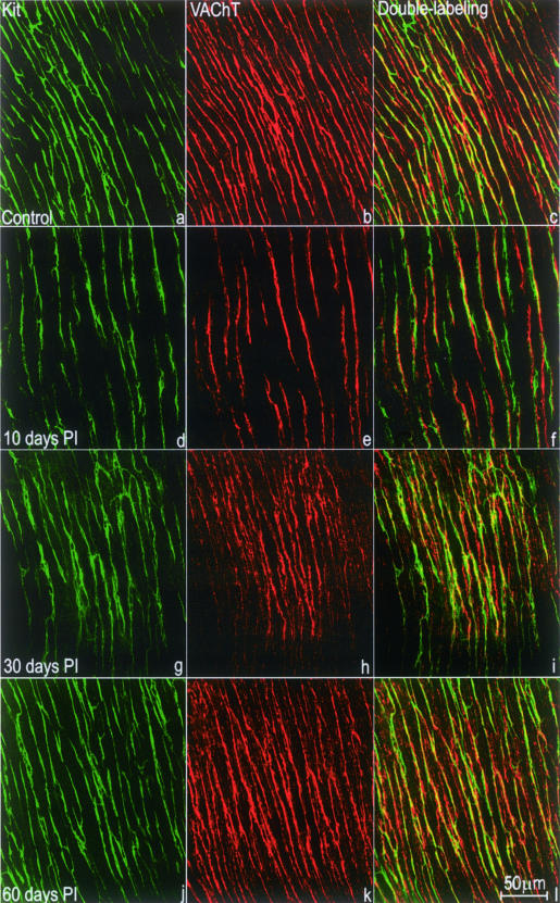
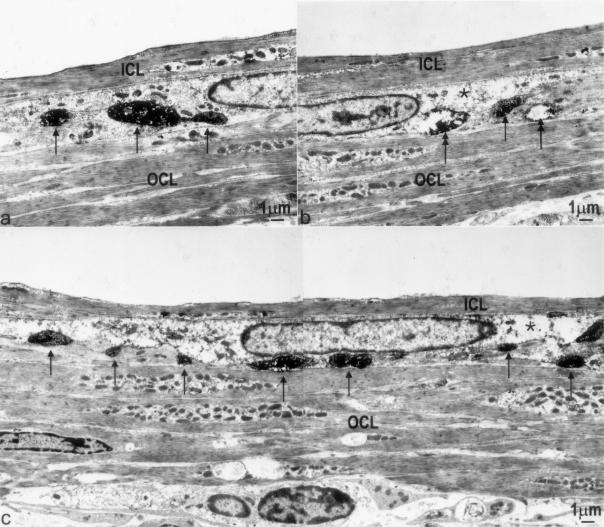
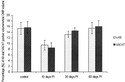
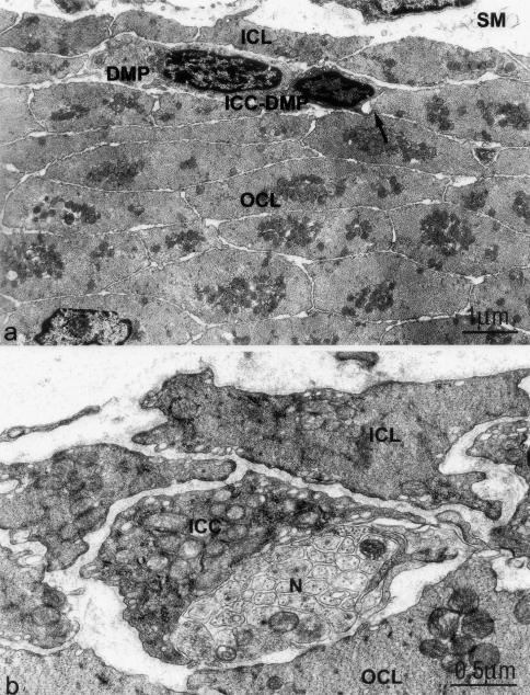
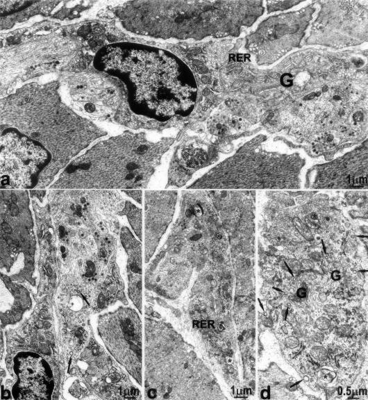
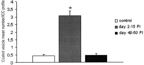
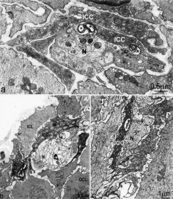
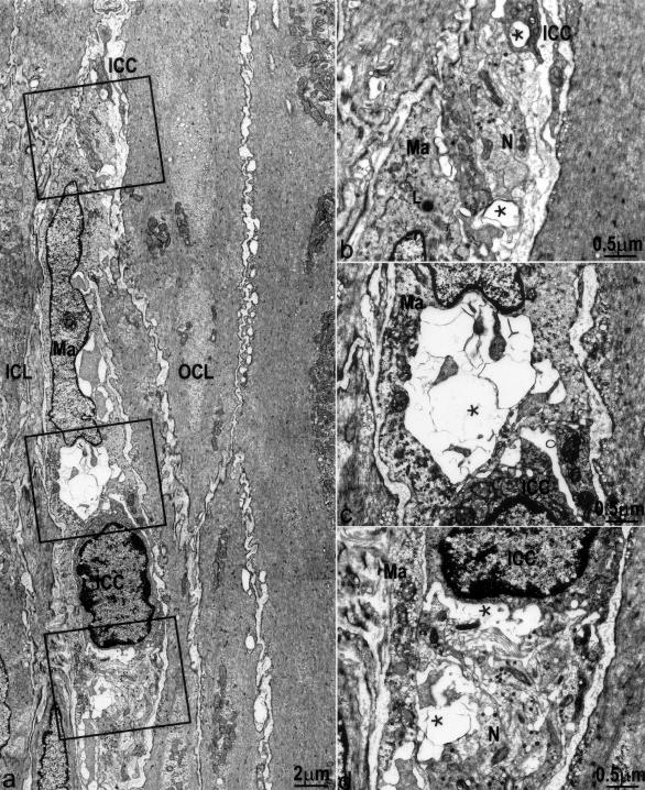
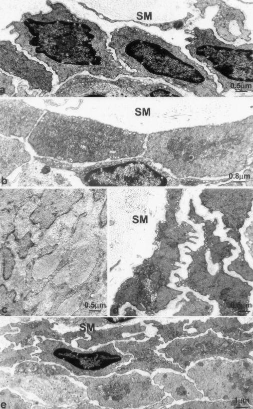
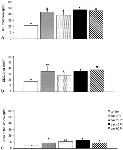
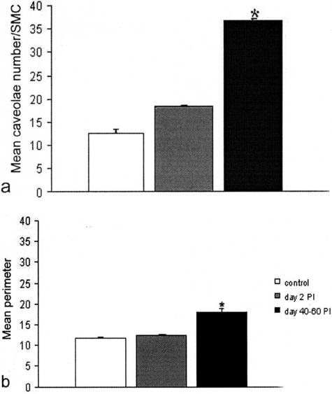
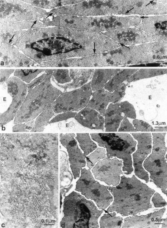
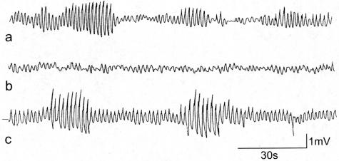
The physiology and pathophysiology of the network of interstitial cells of Cajal associated with the deep muscular plexus (ICC-DMP) of the small intestine are still poorly understood. The objectives of the present study were to evaluate the effects of inflammation associated with Trichinella spiralis infection on the ICC-DMP and to correlate loss of function with structural changes in these cells and associated structures. We used immunohistochemistry, electron microscopy, and assessment of distention-inducing electrophysiological parameters in vitro. Damage to ICC-DMP was associated with a loss of distention-induced patterns of electrical activity normally associated with distention-induced peristalsis. Consistently, the timing of recovery of ICC-DMP paralleled the timing of recovery of the distention-induced activity. Nerve varicosities associated with ICC-DMP including cholinergic nerves, assessed by immunoelectron microscopy and whole mount double labeling, paralleled injury to ICC-DMP thus contributing to impaired excitatory innervation of smooth muscle cells. Major additional changes included a remodeling of the inner circular muscle layer, which may affect long-term sensitivity to distention after infection. In conclusion, transient injury to ICC-DMP in response to T. spiralis infection is severe and associated with a complete lack of distention-induced burst-type muscle activity.


Figures

Figure 1
Figure 1
Whole mount preparations showing immunoreactivities for c-Kit and VAChT at the level of the DMP. a–c: Control; d–f: 10 days after infection; g–i: 30 days after infection; and j–l: 60 days after infection. c-Kit-positive ICC-DMP (green; a, d, g, j) were mostly bipolar in shape and closely apposed to VAChT-positive nerve varicosities (red; b, e, h, k) in the DMP. c, f, i, and l: c-Kit and VAChT double labeling. a to f: At day 10 after infection (d–f), immunoreactivities for both c-Kit and VAChT were reduced compared with control tissue (a–c). g to l: c-Kit and VAChT reactivities recovered to some degree by day 30 after infection (g–i) and returned to normal by day 60 after infection (j–l).
Figure 2
Figure 2
Injury, loss, and recovery of VAChT nerve varicosities. a: Immunoelectron microscopy for VAChT showed regular appearance of cholinergic nerve varicosities (arrows) in the DMP of control tissue. Immunoreactivity is seen as dark area with higher electron density (arrows). b: Ten days after infection, cholinergic varicosities were difficult to find; those present showed (partially) depleted cytoplasm (double arrows). The single arrow indicates an intact nerve varicosity. c: Thirty days after infection, more VAChT-positive nerves (arrows) were observed compared with 10 days after infection, significant ultrastructural damage was still present in the ICC-DMP. *, Partially depleted cytoplasm. ICL, inner circular muscle layer; OCL, outer circular muscle layer.
Figure 3
Figure 3
Quantification of c-Kit and VAChT immunoreactivity. Percentage of c-Kit- and VAChT-positive volume at the level of the DMP (mean ± SE, n = 10), expressed as percent volume of total volume of DMP area in the field. Ten days after infection: significantly reduced compared to control (P < 0.05 for c-Kit reaction; P < 0.01 for VAChT reaction). Thirty days after infection: no significant difference from control (P = 0.38 for c-Kit reaction; P = 0.71 for VAChT reaction). Sixty days after infection: no significant difference from control (P = 0.97 for c-Kit reaction; P = 0.91 for VAChT reaction).
Figure 4
Figure 4
ICC-DMP have close connections with smooth muscle cells and nerve varicosities of the DMP in control tissue. a: An ICC-DMP in the DMP between the inner (ICL) and outer circular muscle layer (OCL). Gap junctions are numerous between smooth muscle cells and between ICC-DMP and smooth muscle cells (arrow). SM, submucosa. b: Numerous mitochondria, endoplasmic reticulum, and caveoli are present in the ICC-DMP. Close connections occur between ICC-DMP and enteric nerve varicosities (N) in the DMP.
Figure 5
Figure 5
Ultrastructural changes in ICC-DMP, 2 to 15 days after infection. a and b: Two days after infection: increased rough endoplasmic reticulum (RER), large Golgi apparatus (G), and coated vesicles (arrows) in ICC-DMP. c: Fifteen days after infection: RER and lamellar bodies (asterisks) in a process of ICC-DMP. d: Fifteen days after infection: coated vesicles (arrows) at the budding face of the Golgi apparatus (G) and close to plasma membrane. *, Lamellar bodies.
Figure 6
Figure 6
Dramatic transient increase in coated vesicles in ICC-DMP. Difference is significant between control and 40 to 60 days after infection versus 2 to 15 days after infection. *P ≤ 0.01. No significant difference between control and 40 to 60 days after infection.
Figure 7
Figure 7
Loss and recovery of cell to cell contacts. a: Two days after infection, lamellar bodies (arrows) are observed at areas of contact between ICC-DMP and between ICC-DMP and enteric nerve varicosities (N). b: Slightly injured ICC-DMP (ICC) and associated nerve structure (N) at day 30 after infection. Close connections between ICC-DMP and nerves reappeared. There are still lamellar bodies (*) within both ICC-DMP and nerve varicosities. c: Fibroblasts, which are also interstitial cells of the DMP, showed much less damage compared to ICC-DMP. Injury to an ICC-DMP (ICC) as reflected by the presence of autophagosomes (*) in its cytoplasm; a neighboring fibroblast (FC) is intact.
Figure 8
Figure 8
Structural injury to ICC-DMP and enteric nerves associated with macrophages 10 days after infection. a: Severe injury to ICC-DMP and nerve varicosities, no significant injury to smooth muscle cells. Injured cells were associated with macrophages (Ma). b–d: Enlarged details of a. ICC-DMP (ICC) and nerve varicosities (N) are associated with macrophages (Ma). *, Damaged structures. Close connections between ICC-DMP and nerves as well as gap junctions between ICC-DMP and smooth muscle cells are not present. L, Lysosome within the macrophage.
Figure 9
Figure 9
Ultrastructure of the inner circular muscle layer. a: Control. The inner circular muscle layer consists of one layer of smooth muscle cells. b: Two days after infection. Increased size of the smooth muscle cells and richness in RER and Golgi apparatus. c: Twenty-three days after infection. Enlarged stroma with collagen deposits and smooth muscle cells with numerous branches. d and e: Forty days after infection (d) and 60 days after infection (e). Richness in caveolae and ramified shape of the smooth muscle cells and stratification of the inner circular muscle layer. SM, submucosa.
Figure 10
Figure 10
Quantification of increase in smooth muscle area. a: Total area of the inner circular muscle layer. Significant difference for control versus 2 days after infection, 23 days after infection, 40 and 60 days after infection. No significant difference among the other groups. b: Area occupied by the inner smooth muscle cells. Increase occurred by day 2 up to 60 days after infection. Significant difference for control versus 2 days after infection, 23 days after infection, 40 and 60 days after infection. c: Area occupied by the stroma in the intercellular space. Significant difference for control versus 2 days after infection, 23 days after infection, and 40 and 60 days after infection. *P < 0.01, **P < 0.05.
Figure 11
Figure 11
Quantification of caveolae and SMC perimeter. a: The mean number of caveolae in the smooth muscle cells (SMCs) of the inner circular muscle layer shows a gradual increase up to a factor 2× 40 to 60 days after infection. Significant difference between control as well as 2 to 15 days after infection versus 40 to 60 days after infection (P < 0.01). No significant difference between control and 2 to 15 days after infection. b: The mean perimeter of the SMC of the inner circular muscle layer increased up to 40 to 60 days after infection. Significant difference for both control and 2 to 15 days after infection versus 40 to 60 days after infection (P < 0.01). No significant difference between control and 2 to 15 days after infection.
Figure 12
Figure 12
Ultrastructural changes in the outer circular muscle layer. a: Control. Arrows indicate gap junctions. b: Two days after infection. Wide edematous areas (E) and absence of gap junctions. c: Eight days after infection. Richness in RER in a smooth muscle cell. d: Forty days after infection. Smooth muscle cells were normal and gap junctions were newly present (arrows), but the stroma between the muscle cells is wider than in control.
Figure 13
Figure 13
Distention induced periodic bursts of slow waves with increased amplitude and superimposed action potentials. a: Control conditions. b: Day 10 after infection, distention did not evoke any burst type activity. c: Day 60 after infection. Distention-induced periodic activity was normal. The recordings in a and c were taken after distention-induced activity stabilized, which was 5 minutes after initiation of distention.

Similar articles

Cited by

References

    1. Thuneberg L. Interstitial cells of Cajal: intestinal pacemaker cells? Adv Anat Embryol Cell Biol. 1982;71:1–130. - PubMed
    1. Rumessen JJ, Thuneberg L, Mikkelsen HB. Plexus muscularis profundus and associated interstitial cells. II. Ultrastructural studies of mouse small intestine. Anat Rec. 1982;203:129–146. - PubMed
    1. Rumessen JJ, Thuneberg L. Plexus muscularis profundus and associated interstitial cells. I. Light microscopical studies of mouse small intestine. Anat Rec. 1982;203:115–127. - PubMed
    1. Faussone Pellegrini MS, Cortesini C. Some ultrastructural features of the muscular coat of human small intestine. Acta Anat (Basel) 1983;115:47–68. - PubMed
    1. Kobilo T, Szurszewski JH, Farrugia G, Hanani M. Coupling and innervation patterns of interstitial cells of Cajal in the deep muscular plexus of the guinea-pig. Neurogastroenterol Motil. 2003;15:635–641. - PubMed

Publication types

MeSH terms

Substances

LinkOut - more resources