Comparative Study
. 2005 Aug;167(2):465-73.
doi: 10.1016/S0002-9440(10)62990-2.

The immunomodulatory proteins B7-DC, B7-H2, and B7-H3 are differentially expressed across gestation in the human placenta

Affiliations
Comparative Study

The immunomodulatory proteins B7-DC, B7-H2, and B7-H3 are differentially expressed across gestation in the human placenta

Margaret G Petroff et al. Am J Pathol. 2005 Aug.

Abstract

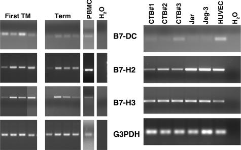
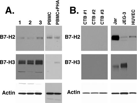
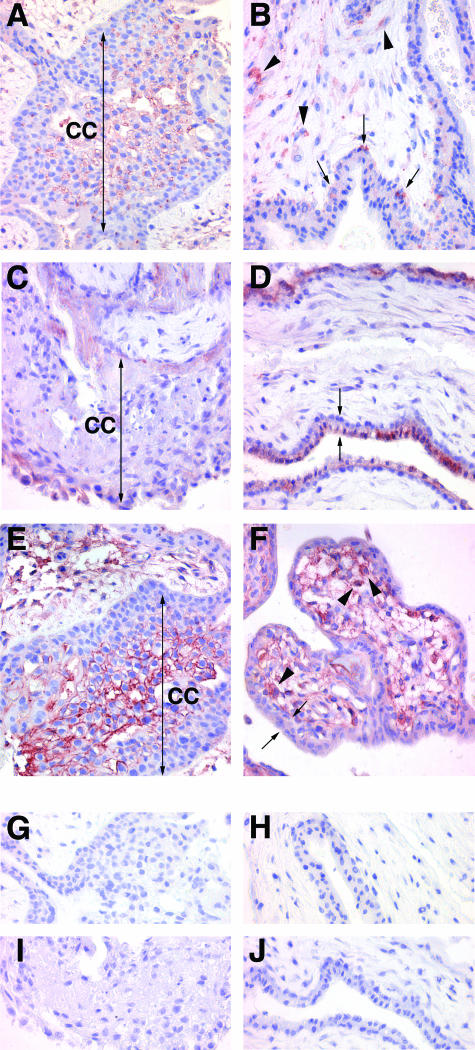
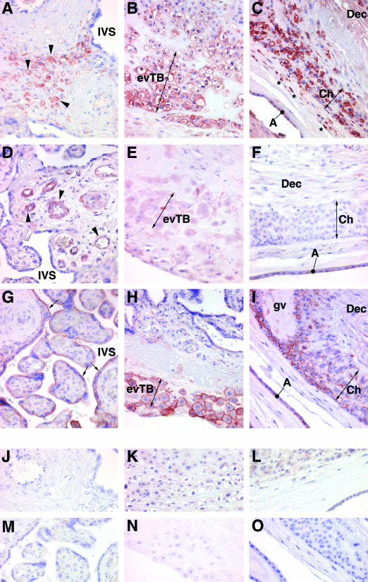
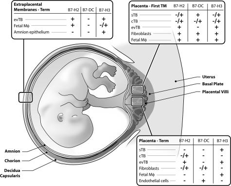
Placental trophoblast cells form a cellular barrier between the potentially immunogenic fetus and maternal leukocytes. Trophoblasts subvert maternal immunity by producing surface-bound and soluble factors that interact with maternal leukocytes. Here, we describe the distribution of three members of the expanding family of B7 immunomodulatory molecules: B7-DC, B7-H2, and B7-H3. B7-DC and B7-H3 inhibit antigen-stimulated lymphocyte activation while B7-H2 serves in a regulatory capacity, often promoting a Th2 immunophenotype. First trimester and term placentas, purified trophoblast cells, choriocarcinoma cell lines, and human umbilical vein endothelial cells were analyzed for B7 family RNA and protein expression. Transcripts and proteins for all three B7s were present throughout gestation but were differentially expressed within the trophoblast and the stroma. Whereas B7-DC was prominent on the syncytiotrophoblast of early placenta, it was absent from the trophoblast at term. In contrast, B7-H2 and B7-H3 were prominent on the extravillous trophoblast throughout gestation. Lastly, stromal cells, including macrophages and endothelial cells, differentially expressed B7-DC, B7-H2, and B7-H3, depending on gestational age. Thus, all three of these newly discovered B7 proteins are differentially positioned at the maternal-fetal interface such that they could steer maternal leukocytes away from a harmful immune response and toward a favorable one.


Figures

Figure 1
Figure 1
Analysis of B7 transcripts in placentas and leukocytes. Ten-μm cryosections of placental tissue were used as a source of RNA so that nearby tissue sections could be counterstained and analyzed microscopically to verify that tissue included solely placental villi. RNA was subjected to RT-PCR using primers specific for B7-DC, B7-H2, B7-H3, or the housekeeping gene, G3PDH. Left: Ethidium bromide-stained agarose gel of RT-PCR products from total RNA of four first trimester placentas, four term placentas, and human PBMCs. H2O: water was substituted for cDNA as a negative control. Right: RT-PCR products of RNA from three different term villous cytotrophoblast preparations (CTB nos. 1, 2, and 3), Jar, JEG-3, and HUVECs.
Figure 2
Figure 2
Immunoblot analysis of term placenta and cells. A: Protein lysates from three different term placentas and from resting and PHA-activated human PBMCs were electrophoresed and subjected to immunoblot analysis and chemiluminescence detection for B7-H2, B7-H3, and actin. B: Protein from three different preparations of term villous cytotrophoblast cell preparations, Jar, JEG-3, or HUVECs were analyzed for these proteins.
Figure 3
Figure 3
Immunohistochemical localization of B7 family proteins in the first trimester placenta. A, C, E, G, and I: Extravillous trophoblast cell columns. B, D, F, H, and J: Villous portions of the placenta. A, B: B7-H2; C, D: B7-DC; E, F: B7-H3; G, H: rabbit IgG control; I, J: goat IgG control. Two-headed arrows in A, C, and E bracket cell columns. Arrowheads in B and F show positively staining stromal cells. Arrows in B show positively stained villous cytotrophoblast cells. Double arrows in D and F bracket the villous cyto- and syncytiotrophoblast. CC, cell column. Original magnifications, ×400.
Figure 4
Figure 4
Immunohistochemical localization of B7 family proteins in the term placenta. A, D, G, J, and M: Villous placenta; B, E, H, K, and N: basal plate placenta; C, F, I, L, and O: amniochorion membrane. AC: B7-H2; D-F: B7-DC; G--I: B7-H3; J--L: rabbit IgG control; MO: goat IgG control. Arrowheads in A and D show positively stained villous stromal cells; two-headed arrows in B, E, H and C, F, I bracket clusters of extravillous trophoblast cells of the basal plate and chorion membrane, respectively; asterisks in C denote positively stained cells within the connective tissue between the amnion and chorion; arrows in G show the syncytiotrophoblast. The amnion membrane in I has been separated from the chorion due to processing artifact, and has doubled back on itself. A, amnion epithelium; Ch, chorion; Dec, decidua; evTB, extravillous trophoblast; IVS intervillous space; gv, ghost villus. Original magnifications, ×400.
Figure 5
Figure 5
Summary of immunohistochemical results in first trimester and term placentas and extraplacental membranes. +, Presence of the protein; +/−, variable presence of the protein; −, the absence of the protein. cTB, cytotrophoblast; evTB, extravillous trophoblast; IVS, intervillous space; Mϕ, macrophages; sTB, syncytiotrophoblast.

References

    1. Petroff MG, Hunt JS. Immunity at the maternal-fetal interface. Mestecky J, Bienenstock J, Lamm ME, Mayer L, McGhee JR, Strober W, editors. San Diego: Elsevier, Inc.,; Mucosal Immunology. 2005
    1. June CH, Bluestone JA, Nadler LM, Thompson CB. The B7 and CD28 receptor families. Immunol Today. 1994;15:321–331. - PubMed
    1. Greenfield EA, Nguyen KA, Kuchroo VK. CD28/B7 costimulation: a review. Crit Rev Immunol. 1998;18:389–418. - PubMed
    1. Petroff MG, Chen L, Phillips TA, Azzola D, Sedlmayr P, Hunt JS. B7 family molecules are favorably positioned at the human maternal-fetal interface. Biol Reprod. 2003;68:1496–1504. - PubMed
    1. Nishimura H, Minato N, Nakano T, Honjo T. Immunological studies on PD-1-deficient mice: implication of PD-1 as a negative regulator for B cell responses. Int Immunol. 1998;10:1563–1572. - PubMed

Publication types

MeSH terms