. 2005 Aug;167(2):475-88.
doi: 10.1016/S0002-9440(10)62991-4.

Stroma-derived three-dimensional matrices are necessary and sufficient to promote desmoplastic differentiation of normal fibroblasts

Affiliations

Stroma-derived three-dimensional matrices are necessary and sufficient to promote desmoplastic differentiation of normal fibroblasts

Michael D Amatangelo et al. Am J Pathol. 2005 Aug.

Abstract

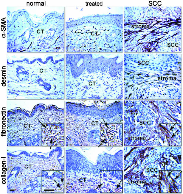
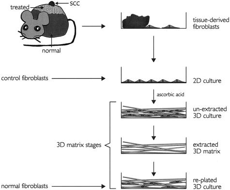
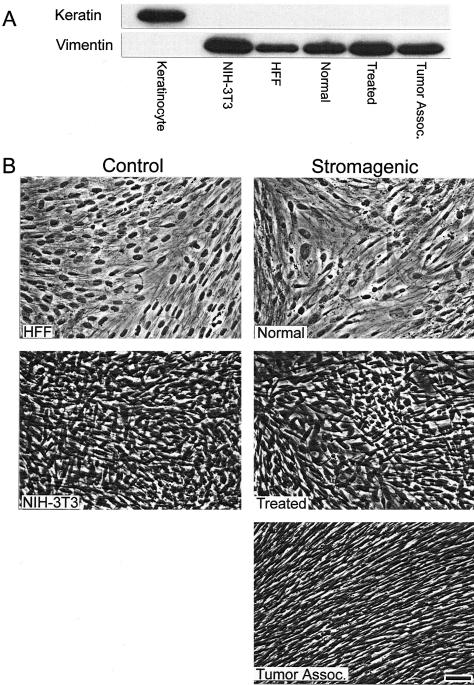
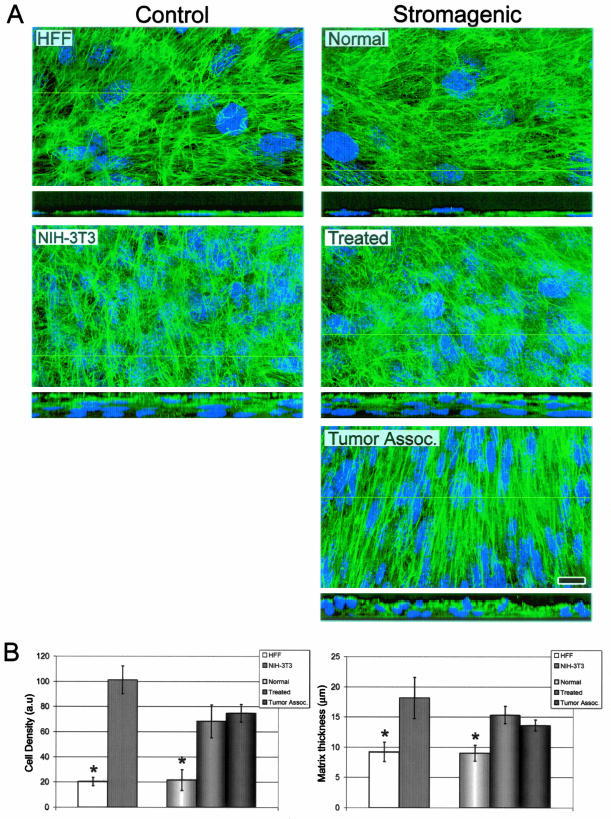
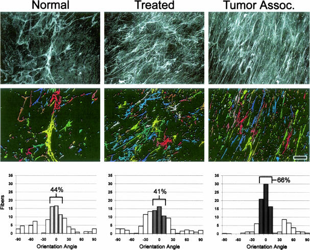
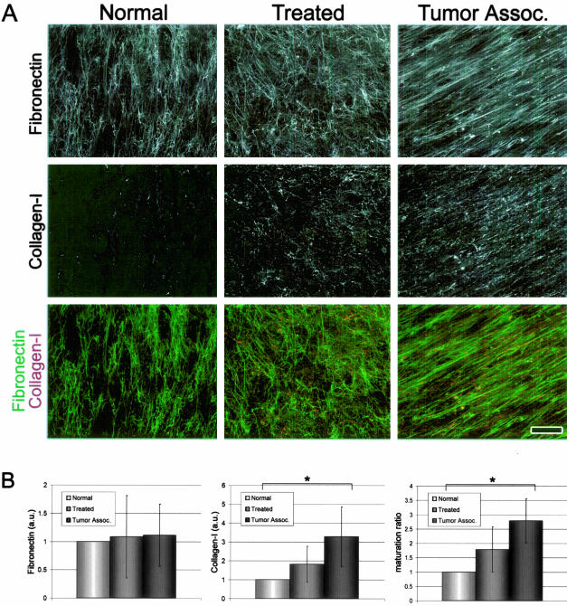
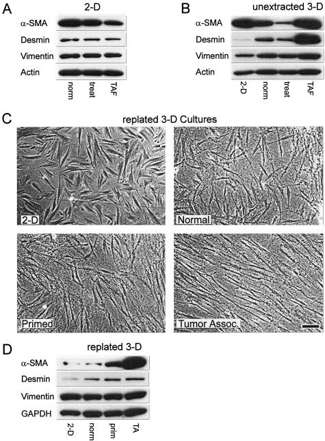
Stromagenesis is a host reaction of connective tissue that, when induced in cancer, produces a progressive and permissive mesenchymal microenvironment, thereby supporting tumor progression. The stromal microenvironment is complex and comprises several cell types, including fibroblasts, the primary producers of the noncellular scaffolds known as extracellular matrices. The events that support tumor progression during stromagenesis are for the most part unknown due to the lack of suitable, physiologically relevant, experimental model systems. In this report, we introduce a novel in vivo-like three-dimensional system derived from tumor-associated fibroblasts at diverse stages of tumor development that mimic the stromagenic features of fibroblasts and their matrices observed in vivo. Harvested primary stromal fibroblasts, obtained from different stages of tumor development, did not retain in vivo stromagenic characteristics when cultured on traditional two-dimensional substrates. However, they were capable of effectively maintaining the tumor-associated stromal characteristics within three-dimensional cultures. In this study, we demonstrate that in vivo-like three-dimensional matrices appear to have the necessary topographical and molecular information sufficient to induce desmoplastic stroma differentiation of normal fibroblasts.


Figures

Figure 1
Figure 1
SCC stroma exhibits a desmoplastic phenotype in vivo. Immunohistochemistry counterstained with hematoxylin; Gill’s no. 3 nuclei stain, of normal skin (normal), treated skin (treated), and SCC showing α-SMA, desmin, fibronectin, and type I collagen. Connective tissue (CT), tumor-associated stroma (stroma), and SCC are denoted. Arrows identify ECM fibers. Note the levels of expression of α-SMA and desmin within the tumor-associated stroma. Also note the disorganization of the ECM fibers of both fibronectin and collagen within normal and treated CT, depicted by the insets versus the parallel patterned ECM organization within the chemically induced mouse SCC-associated stroma. Scale bars, 30 μm.
Figure 2
Figure 2
Model illustrating production and staging of fibroblast-derived 3-D matrices. Mouse skin SCCs were induced by a two-step process (see Materials and Methods). Indicated (normal, treated, and SCC) tissue samples were placed onto tissue culture plates where fibroblasts were allowed to crawl out. Tissue samples were removed and cells were stored or allowed to reach confluence for experimental purposes (2-D culture). Stromagenic and control 2-D cultures were kept for 8 days under conditions permissive for matrix production (unextracted 3-D cultures). Unextracted cultures were used for experimental purposes or submitted to alkaline detergent extraction (extracted 3-D matrices). Extracted 3-D matrices were stored at 4°C until needed for experimental use. Finally HFFs (normal fibroblasts) were replated into extracted 3-D matrices (replated 3-D culture). Note that normal, treated, or TAFs, as well as control fibroblasts, were used to form all three unextracted, extracted, and replated 3-D matrix stages.
Figure 3
Figure 3
Unextracted control and stromagenic 3-D cultures are fibroblastic and homogenous. A: Western blot showing the expression of epithelial marker keratin-10 and fibroblastic marker vimentin. Control cells were epithelial primary mouse keratinocytes and fibroblastic HFFs and NIH-3T3. Stromagenic normal, treated, and tumor-associated cells were positive for vimentin and negative for keratin expression. B: Phase contrast images of unextracted fibroblastic cultures showing morphological differences between stromagenic cell types and control fibroblasts. Note organizational level of tumor-associated unextracted 3-D cultures and similarities between HFF and normal and between NIH-3T3 and treated unextracted 3-D cultures in appearance, as well as the overcoming of growth inhibition by contact of treated and tumor-associated cultures. Scale bar, 25 μm.
Figure 4
Figure 4
Normal unextracted 3-D cultures reach cell densities and matrix thicknesses that significantly differ from stromagenic-treated and tumor-associated unextracted cultures. A: Maximum projection reconstructed confocal images of indirect immunofluorescently labeled fibronectin (in green) and cell nuclei (in blue) of assorted control and stromagenic unextracted 3-D cultures. The yellow lines in the main panels indicate the position where insets, at the bottom of the main panels, show 90° views of the unextracted cultures. Note, within the insets, that normal-derived monolayers and stromagenic-derived polylayers vary in matrix thicknesses. B: Bar graphs representing the average measured cell density and matrix thickness for all assorted cell types (see Table 1 for details). Asterisks indicate statistical significant differences from all other data in the graphs. Scale bar, 15 μm.
Figure 5
Figure 5
Stromagenic fibronectin 3-D fibers are progressively organized in parallel patterns. Deconvoluted maximum projection reconstructed images of indirect immunofluorescently labeled fibronectin fibers of normal, treated, and tumor-associated unextracted 3-D cultures (top). Images were analyzed to create a binary image output that identifies individual fibers by presenting them as variable pseudo-colored objects (middle; see Materials and Methods for details). Angles at which fibers were orientated relative to the x-axis were rounded to the nearest 10th degree and are shown in the graphs (bottom). The sample mode was arbitrarily set to 0°, thus normalizing the data for comparison purposes (see Table 1 for details and statistics). Numbers indicate percentages of angles positioned within 10° of the angle for which most of the fibers were orientated. Note that less than 50% of fibers were found to be organized in normal and primed (treated) matrices, while more than 60% of fibers were found to be organized parallel within tumor-associated 3-D matrices. Scale bar, 15 μm.
Figure 6
Figure 6
The maturation state of 3-D matrices intensifies in a stromagenic-dependent manner. A: Maximum projection reconstructed confocal images of indirect immunofluorescence showing ECM protein assembly in stromagenic unextracted 3-D cultures. Samples were labeled for fibronectin (top) or type I collagen (middle). Overlaid images showing fibronectin (in green) and type I collagen (collagen-I, in red) are shown at the bottom. Images were submitted to identical filters (see Materials and Methods for details). B: Graph bar representing quantification of fibronectin fibers, type I collagen fibers, and matrix maturation ratio for each stromagenic cell type (see Table 1 for details and statistics). Asterisks mark samples statistically significantly different from all others. Note the increase in type I collagen levels and 3-D matrix maturation during stromagenesis. Scale bar, 25 μm.
Figure 7
Figure 7
Extracted 3-D matrices derived from assorted stromagenic phases differentially modify primary normal fibroblasts. Western blots showing protein levels of α-SMA, desmin, and vimentin expression on 2-D (A) or within pre-extracted 3-D (B) cultures for normal (norm), treated (treat), or TAFs. C: Phase contrast images of normal primary human fibroblasts plated on 2-D coated fibronectin or within normal, primed, or tumor-associated extracted 3-D matrices. D: Western blot showing α-SMA, desmin, and vimentin expression levels of normal primary human fibroblasts plated on 2-D coated fibronectin or within normal (norm), primed (prim), or tumor-associated (TA) extracted 3-D matrices (see Table 1 for statistics). Note how different stromagenic phases differentially modify the normal primary fibroblast morphology, organization, and desmoplastic marker expression. Scale bar, 25 μm.

References

    1. Bissell MJ, Radisky D. Putting tumours in context. Nat Rev Cancer. 2001;1:46–54. - PMC - PubMed
    1. Kunz-Schughart LA, Knuechel R. Tumor-associated fibroblasts (part I): active stromal participants in tumor development and progression? Histol Histopathol. 2002;17:599–621. - PubMed
    1. Kunz-Schughart LA, Knuechel R. Tumor-associated fibroblasts (part II): functional impact on tumor tissue. Histol Histopathol. 2002;17:623–637. - PubMed
    1. Silzle T, Randolph GJ, Kreutz M, Kunz-Schughart LA. The fibroblast: sentinel cell and local immune modulator in tumor tissue. Int J Cancer. 2004;108:173–180. - PubMed
    1. Mueller MM, Fusenig NE. Friends or foes—bipolar effects of the tumour stroma in cancer. Nat Rev Cancer. 2004;4:839–849. - PubMed

MeSH terms

Substances