. 2005 Aug;167(2):505-15.
doi: 10.1016/s0002-9440(10)62993-8.

Cerebral microvascular amyloid beta protein deposition induces vascular degeneration and neuroinflammation in transgenic mice expressing human vasculotropic mutant amyloid beta precursor protein

Affiliations

Cerebral microvascular amyloid beta protein deposition induces vascular degeneration and neuroinflammation in transgenic mice expressing human vasculotropic mutant amyloid beta precursor protein

Jianting Miao et al. Am J Pathol. 2005 Aug.

Abstract

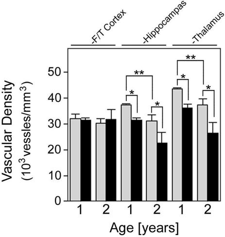
Cerebral vascular amyloid beta-protein (Abeta) deposition, also known as cerebral amyloid angiopathy, is a common pathological feature of Alzheimer's disease. Additionally, several familial forms of cerebral amyloid angiopathy exist including the Dutch (E22Q) and Iowa (D23N) mutations of Abeta. Increasing evidence has associated cerebral microvascular amyloid deposition with neuroinflammation and dementia in these disorders. We recently established a transgenic mouse model (Tg-SwDI) that expresses human vasculotropic Dutch/Iowa mutant amyloid beta-protein precursor in brain. Tg-SwDI mice were shown to develop early-onset deposition of Abeta exhibiting high association with cerebral microvessels. Here we present quantitative temporal analysis showing robust and progressive accumulation of cerebral microvascular fibrillar Abeta accompanied by decreased cerebral vascular densities, the presence of apoptotic cerebral vascular cells, and cerebral vascular cell loss in Tg-SwDI mice. Abundant neuroinflammatory reactive astrocytes and activated microglia strongly associated with the cerebral microvascular fibrillar Abeta deposits. In addition, Tg-SwDI mouse brain exhibited elevated levels of the inflammatory cytokines interleukin-1beta and -6. Together, these studies identify the Tg-SwDI mouse as a unique model to investigate selective accumulation of cerebral microvascular amyloid and the associated neuroinflammation.


Figures

Figure 1
Figure 1
Fibrillar Aβ deposits develop exclusively in the cerebral microvasculature of Tg-SwDI mice. Immunolabeling for Aβ (A) and negative thioflavin S staining (B and C) in the neocortex of 12-month-old Tg-SwDI mice indicates that the abundant diffuse parenchymal deposits in this region are not fibrillar. Immunolabeling for Aβ (D) and thioflavin S staining (E) in the thalamus co-localize (F) identifying abundant microvascular fibrillar amyloid deposits in this region of Tg-SwDI mice. Immunolabeling for collagen IV (G) and thioflavin S staining (H) in the thalamus co-localize (I) confirming the microvascular localization of the fibrillar amyloid in the thalamus of Tg-SwDI mice. Scale bars, 50 μm.
Figure 2
Figure 2
Distribution of Aβ isoforms in parenchymal diffuse and cerebral vascular fibrillar deposits in Tg-SwDI mice. Immunostaining of 12-month-old Tg-SwDI mouse brain sections for collagen IV (red) and Aβ40 or Aβ42 (brown) in the fronto-temporal cortex or thalamus. The diffuse parenchymal deposits showed stronger immunostaining for Aβ40 than Aβ42 (A and B, respectively) whereas the fibrillar microvascular deposits in the thalamus exhibited strong staining for Aβ40 and Aβ42 (C and D, respectively). E: ELISA analysis for total Aβ40 and Aβ42 in microvessel and nonvascular parenchymal fractions isolated from 12-month-old Tg-SwDI mice. Data shown are the mean ± SD (n = 4 mice). *P < 0.0002 and **P < 0.0001.
Figure 3
Figure 3
Progressive cerebral microvascular Aβ accumulation in Tg-SwDI mice. A: Representative thalamic region from a 12-month-old Tg-SwDI mouse was immunostained for Aβ (brown) and collagen IV (red). B: Quantitative stereological measurement of microvascular Aβ deposition in different brain regions of increasing aged Tg-SwDI mice. Data shown are the mean ± SD (n = 4 mice for each age group). Scale bar, 100 μm.
Figure 4
Figure 4
Meningeal vessel Aβ accumulation in Tg-SwDI mice. Immunostaining for Aβ (A) and thioflavin S staining (B) co-localize (C) demonstrating fibrillar amyloid deposition in a meningeal vessel of Tg-SwDI mice. D: Quantitative stereological measurement of meningeal vessel Aβ deposition of increasing aged Tg-SwDI mice. Data shown are the mean ± SD (n = 4 mice for each age group). Scale bars, 50 μm.
Figure 5
Figure 5
Decreased regional vascular densities in Tg-SwDI mice. Vascular densities were determined in different brain regions of 1- and 2-year-old wild-type mice (gray bars) and Tg-SwDI mice (black bars) as described in Materials and Methods. Data shown are the mean ± SD (n = 4 mice for each group). *P < 0.01 and **P < 0.02.
Figure 6
Figure 6
Apoptotic cerebral vascular cells in Tg-SwDI mice. Immunolabeling for Aβ (A) or collagen IV (B and C) in red and TUNEL labeling in green identified apoptotic vascular cells in meningeal vessels (A and B) and thalamic microvessels (C) in 2-year-old Tg-SwDI mice. Scale bar, 50 μm.
Figure 7
Figure 7
Loss of smooth muscle cells in amyloid-laden meningeal vessels of Tg-SwDI mice. Immunolabeling for Aβ (A) and vascular smooth muscle cell α-actin (B) in a meningeal vessel of a 2-year-old Tg-SwDI mouse. The arrowheads reveal loss of smooth muscle cells in the amyloid-rich regions of the vessel as shown in the merge image (C). Scale bar, 50 μm.
Figure 8
Figure 8
Ultrastructural analysis of cerebral microvascular amyloid deposition in Tg-SwDI mice. A: Normal thalamic microvessel in a wild-type mouse. Vessel lumen (L) and vascular basement membrane (*) are identified. B: Thalamic microvessel in a Tg-SwDI mouse with an amyloid deposit (A) and degenerating pericytes with swollen vacuoles (white arrowheads). C: Tg-SwDI cerebral microvessel with amyloid deposition and an engaged microglial cell (M). Initial penetrance of amyloid into the vascular basement membrane is identified by the open arrowhead. D: Another thalamic microvessel in a Tg-SwDI mouse with more extensive amyloid deposition extending into the surrounding parenchyma, several sites of amyloid breaching the vascular basement membrane, and several engaged microglial cells. Scale bar, 2.5 μm.
Figure 9
Figure 9
Increased cerebral microvascular-associated reactive astrocytes in Tg-SwDI mice. Immunostaining for GFAP (brown) and collagen IV (red) for identifying astrocytes and microvessels, respectively, in different brain regions of 1-year-old wild-type mice (A–C) and Tg-SwDI mice (D–F). G: Quantitative stereological measurement of astrocyte densities in brain regions of increasing aged wild-type mice (gray bars) and Tg-SwDI mice (black bars). Data shown are mean ± SD (n = 4 mice for each group). Scale bar, 50 μm.
Figure 10
Figure 10
Increased cerebral microvascular-associated activated microglia in Tg-SwDI mice. Immunostaining for mAb 5D4 (brown) and collagen IV (red) for identifying activated microglia and microvessels, respectively, in different brain regions of 1-year-old wild-type mice (A–C) and Tg-SwDI mice (D–F). G: Quantitative stereological measurement of activated microglial densities in brain regions of increasing aged Tg-SwDI mice. Data shown are mean ± SD (n = 4 mice for each group). Scale bar, 50 μm.
Figure 11
Figure 11
Elevated levels of inflammatory cytokines in Tg-SwDI mouse brain. The levels of IL-1β (gray bars) and IL-6 (black bars) in soluble brain extracts prepared from 12-month-old wild-type and Tg-SwDI mice were determined by ELISA analysis as described in Materials and Methods. Data shown are mean ± SD (n = 5 mice for each group). *P < 0.0001.

References

    1. Selkoe DJ. Alzheimer’s disease: genes, proteins, and therapy. Physiol Rev. 2001;81:741–766. - PubMed
    1. Jellinger KA. Alzheimer’s disease and cerebrovascular pathology: an update. J Neural Transm. 2002;109:813–836. - PubMed
    1. Vinters HV, Farag ES. Amyloidosis of cerebral arteries. Adv Neurol. 2003;92:105–112. - PubMed
    1. Selkoe DJ. Deciphering the genesis and fate of amyloid beta-protein yields novel therapies for Alzheimer disease. J Clin Invest. 2002;110:1375–1381. - PMC - PubMed
    1. Levy E, Carman MD, Fernandez-Madrid IJ, Power MD, Lieberburg I, van Duinen SG, Bots GTAM, Luyendijk W, Frangione B. Mutation of the Alzheimer’s disease amyloid gene in hereditary cerebral hemorrhage, Dutch type. Science. 1990;248:1124–1126. - PubMed

Publication types

MeSH terms