

Human cord blood stem cells generate human cytokeratin 18-negative hepatocyte-like cells in injured mouse liver
- PMID: 16049339
- PMCID: PMC1603572
- DOI: 10.1016/S0002-9440(10)62997-5
Human cord blood stem cells generate human cytokeratin 18-negative hepatocyte-like cells in injured mouse liver
Abstract
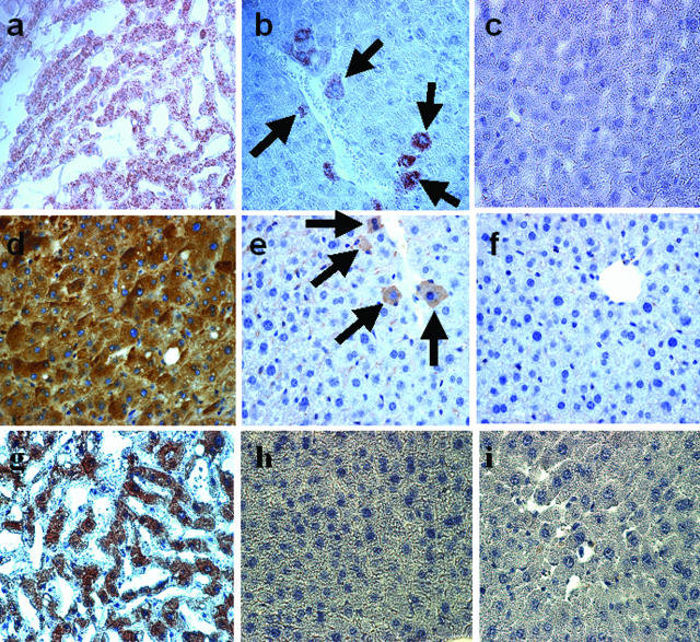
Differentiation of adult bone marrow (BM) cells into nonhematopoietic cells is a rare phenomenon. Several reports, however, suggest that human umbilical cord blood (hUCB)-derived cells give rise to hepatocytes after transplantation into nonobese diabetic-severe combined immunodeficient (NOD-SCID) mice. Therefore, we analyzed the hepatic differentiation potential of hUCB cells and compared the frequency of newly formed hepatocyte-like cells in the livers of recipient NOD-SCID mice after transplantation of hUCB versus murine BM cells. Mononuclear cell preparations of hUCB cells or murine BM from enhanced green fluorescent protein transgenic or wild-type mice were transplanted into sublethally irradiated NOD-SCID mice. Liver regeneration was induced by carbon tetrachloride injury with and without subsequent hepatocyte growth factor treatment. By immunohistochemistry and reverse transcriptase-polymerase chain reaction, we detected clusters of hepatocyte-like cells in the livers of hUCB-transplanted mice. These cells expressed human albumin and Hep Par 1 but mouse CK18, suggesting the formation of chimeric hepatocyte-like cells. Native fluorescence microscopy and double immunofluorescence failed to detect single hepatocytes derived from transplanted enhanced green fluorescent protein-transgenic mouse BM. Fluorescent in situ hybridization rarely revealed donor-derived hepatocyte-like cells after cross-gender mouse BM transplantation. Thus, hUCB cells have differentiation capabilities different from murine BM cells after transplantation into NOD-SCID mice, demonstrating the importance of further testing before hUCB cells can be used therapeutically.
Figures


Similar articles
-
Human umbilical cord blood-derived cells differentiate into hepatocyte-like cells in the Fas-mediated liver injury model.Am J Physiol Gastrointest Liver Physiol. 2005 Dec;289(6):G1091-9. doi: 10.1152/ajpgi.00049.2005. Epub 2005 Jul 28. Am J Physiol Gastrointest Liver Physiol. 2005. PMID: 16051923
-
In vivo hepatic differentiation of mesenchymal stem cells from human umbilical cord blood after transplantation into mice with liver injury.Biochem Biophys Res Commun. 2012 Jun 15;422(4):539-45. doi: 10.1016/j.bbrc.2012.04.156. Epub 2012 May 9. Biochem Biophys Res Commun. 2012. PMID: 22580002
-
Intrahepatic transplantation of CD34+ cord blood stem cells into newborn and adult NOD/SCID mice induce differential organ engraftment.Tissue Cell. 2012 Apr;44(2):80-6. doi: 10.1016/j.tice.2011.11.004. Epub 2011 Dec 23. Tissue Cell. 2012. PMID: 22197619
-
Immune-deficient mouse models for analysis of human stem cells.Biotechniques. 2003 Dec;35(6):1262-72. doi: 10.2144/03356ss06. Biotechniques. 2003. PMID: 14682062 Review.
-
Contribution of human hematopoietic stem cells to liver repair.Semin Immunopathol. 2009 Sep;31(3):411-9. doi: 10.1007/s00281-009-0166-3. Epub 2009 Jun 17. Semin Immunopathol. 2009. PMID: 19533133 Free PMC article. Review.
Cited by
-
Therapeutic Potential of HGF-Expressing Human Umbilical Cord Mesenchymal Stem Cells in Mice with Acute Liver Failure.Int J Hepatol. 2016;2016:5452487. doi: 10.1155/2016/5452487. Epub 2016 Feb 28. Int J Hepatol. 2016. PMID: 27057357 Free PMC article.
-
Alveolar epithelial cell therapy with human cord blood-derived hematopoietic progenitor cells.Am J Pathol. 2011 Mar;178(3):1329-39. doi: 10.1016/j.ajpath.2010.11.062. Am J Pathol. 2011. PMID: 21356383 Free PMC article.
-
Primary Hepatocyte Isolation and Cultures: Technical Aspects, Challenges and Advancements.Bioengineering (Basel). 2023 Jan 18;10(2):131. doi: 10.3390/bioengineering10020131. Bioengineering (Basel). 2023. PMID: 36829625 Free PMC article. Review.
-
Persistence of a chimerical phenotype after hepatocyte differentiation of human bone marrow mesenchymal stem cells.Cell Prolif. 2008 Feb;41(1):36-58. doi: 10.1111/j.1365-2184.2007.00507.x. Cell Prolif. 2008. PMID: 18211285 Free PMC article.
-
The role of stem cells in physiology, pathophysiology, and therapy of the liver.Stem Cell Rev. 2006;2(1):51-8. doi: 10.1007/s12015-006-0009-8. Stem Cell Rev. 2006. PMID: 17142887 Review.
References
-
- Petersen BE, Bowen WC, Patrene KD, Mars WM, Sullivan AK, Murase N, Boggs SS, Greenberger JS, Goff JP. Bone marrow as a potential source of hepatic oval cells. Science. 1999;284:1168–1170. - PubMed
-
- Alison MR, Poulsom R, Jeffrey R, Dhillon AP, Qualglia A, Jacob J, Novelli M, Prentice G, Williamson J, Wright NA. Hepatocytes from non-hepatic adult stem cells. Nature. 2000;406:257. - PubMed
-
- Krause DS, Theise ND, Collector MI, Henegariu O, Hwang S, Gardner R, Neutzel S, Sharkis SJ. Multi-organ, multi-lineage engraftment by a single bone marrow-derived stem cell. Cell. 2001;105:369–377. - PubMed
-
- Jang YY, Collector MI, Baylin SB, Diehl AM, Sharkis SJ. Hematopoietic stem cells convert into liver cells within days without fusion. Nat Cell Biol. 2004;6:532–539. - PubMed
-
- Harris RG, Herzog EL, Bruscia EM, Grove JE, Van Arnam JS, Krause DS. Lack of a fusion requirement for development of bone marrow-derived epithelia. Science. 2004;305:90–93. - PubMed
Publication types
MeSH terms
Substances
LinkOut - more resources
Full Text Sources
Medical
