Comparative Study
. 2005 Aug;167(2):577-84.
doi: 10.1016/S0002-9440(10)62999-9.

Expression of tight-junction protein claudin-7 is an early event in gastric tumorigenesis

Affiliations
Comparative Study

Expression of tight-junction protein claudin-7 is an early event in gastric tumorigenesis

Adam H Johnson et al. Am J Pathol. 2005 Aug.

Abstract

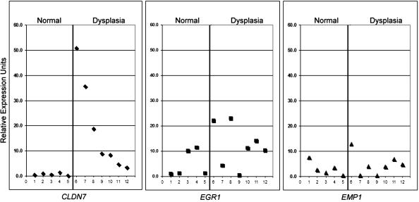
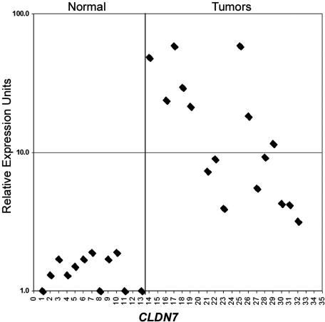
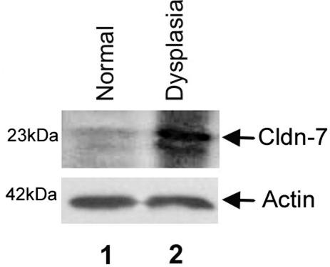
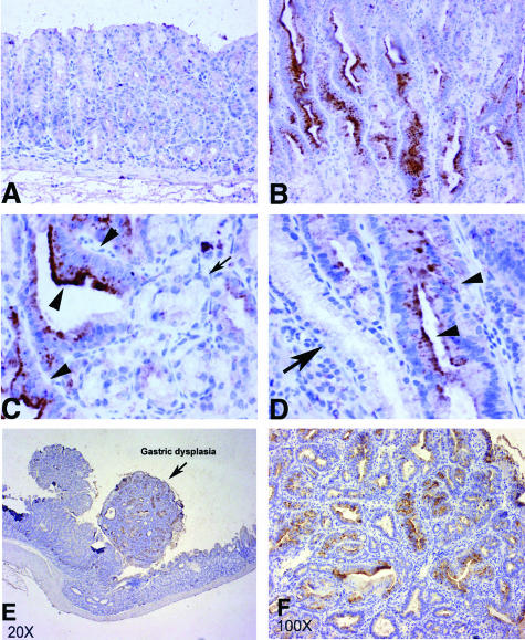
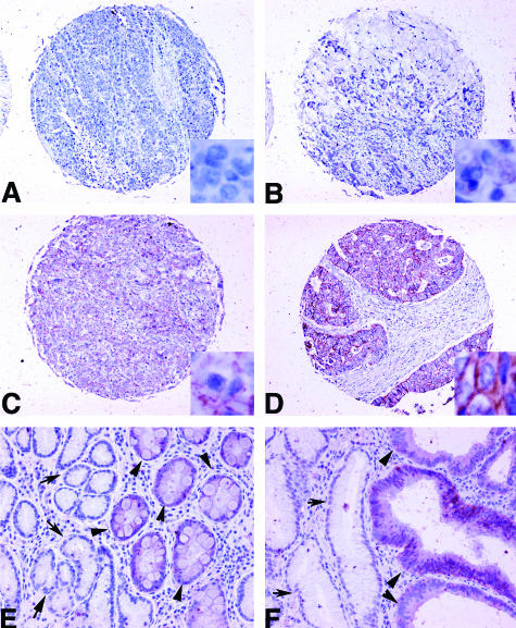
Trefoil factor-1 (Tff1) expression is remarkably down-regulated in nearly all human gastric cancers. Therefore, we used the Tff1 knockout mouse model to detect molecular changes in preneoplastic gastric dysplasia. Oligonucleotide microarray gene expression analysis of gastric dysplasia of Tff1-/- mice was compared to that of normal gastric mucosa of wild-type mice. The genes most overexpressed in Tff1-/- mice included claudin-7 (CLDN7), early growth response-1 (EGR1), and epithelial membrane protein-1 (EMP1). Quantitative real-time reverse transcriptase-polymerase chain reaction and immunohistochemistry showed that Cldn7 was overexpressed in all 10 Tff1-/- gastric dysplasia samples. Comparison with our serial analysis of gene expression database of human gastric cancer revealed similar deregulation in human gastric cancers. Quantitative real-time reverse transcriptase-polymerase chain reaction of human gastric adenocarcinoma samples indicated that, of these three genes, CLDN7 was the most frequently up-regulated gene. Using immunohistochemistry, both mouse and human gastric glands overexpressed Cldn7 in dysplastic but not surrounding normal glands. Cldn7 expression was observed in 30% of metaplasia, 80% of dysplasia, and 70% of gastric adenocarcinomas. Interestingly, 82% of human intestinal-type gastric adenocarcinomas expressed Cldn7 whereas diffuse-type gastric adenocarcinomas did not (P < 0.001). These results suggest that Cldn7 expression is an early event in gastric tumorigenesis that is maintained throughout tumor progression.


Figures

Figure 1
Figure 1
Quantitative real-time RT-PCR (iCycle, Bio-Rad) of mouse CLDN7 (left), EGR1 (middle), and EMP1 (right) on seven microdissected gastric dysplastic tissues of Tff1−/− and six normal gastric mucosa of Tff1+/+. The sample numbers are given in the horizontal axis. The average threshold cycle number for all normal samples was used as reference value. All results were normalized to the expression of β-actin in the same sample. Each sample was compared to six normal samples and there was less than 10% variation for all comparisons. The expression of CLDN7 was higher in all dysplastic tissue samples as compared to normal samples.
Figure 2
Figure 2
Quantitative real-time RT-PCR of human CLDN7 on 13 different normal mucosa samples of the stomach (left) and 20 gastric adenocarcinoma (right) samples. The sample number is given in the horizontal axis. For visualization, the relative expression for all samples is shown in a logarithmic scale. The average threshold cycle number for all normal samples was used as reference value. All results were normalized to the expression of HPRT1 in the same sample, which had minimal variation in all normal and neoplastic gastric samples that we tested. The SEM showed less than 10% variation for all comparisons.
Figure 3
Figure 3
Western blot analysis of claudin-7 in normal and dysplastic stomach of the Tff1+/+ and Tff1−/− mice. Protein extracted from normal stomach (antrum) is loaded in lane 1 and protein extracted from the dysplastic lesion (antrum) is loaded in lane 2. Membranes were immunoblotted with the claudin-7 antibody C-15 (Santa Cruz Biotechnology) at 1:100 dilutions (top) or β-actin antibody AC-74 (Sigma-Aldrich) at 1:1000 dilutions for normalization of protein loading (bottom). The normalization with β-actin (∼42 kd), as shown at the bottom, indicates equal protein loading. The dysplastic tissues show a robust signal for cldn7 (∼23 kd) and the normal tissue shows a very weak signal confirming that cldn7 is specifically overexpressed in gastric dysplasia of the Tff1−/− mice.
Figure 4
Figure 4
Immunohistochemical staining using claudin-7 antibody on gastric tissues of four different Tff1−/− mice. A: Normal gastric mucosa shows no staining. B: A dysplastic tissue of the same mouse as in A showing moderate to strong immunostaining of dysplastic tissues. C: A dysplastic gland next to a normal gland in a second mouse showing increased staining in dysplasia and absent staining in normal glands (indicated by arrows). D: Dysplastic gland next to normal glands showing increased staining in dysplasia of a third different mouse. Arrows indicate normal glands and arrowheads indicate dysplastic glands. E: A low magnification of a dysplastic lesion indicated by arrows in a different mouse. Notice the nodular shape and moderate to strong staining. F: Higher magnification of the lesion shown in E, demonstrating intense staining of dysplastic glands. Original magnifications: ×200 (A, B); ×400 (C, D); ×20 (E); ×100 (F).
Figure 5
Figure 5
Immunohistochemical staining using claudin-7 antibody on tissue array of human gastric tissues. A: A gastric adenocarcinoma showing no staining. B: A gastric adenocarcinoma showing weak intensity staining (1+). C: A gastric adenocarcinoma showing moderate intensity staining (2+). D: A gastric adenocarcinoma showing strong intensity staining (3+). E: Metaplasia adjacent to normal gastric mucosa, showing 1+ staining. Arrows indicate the normal glands with no staining. Arrowheads indicate metaplastic glands. F: Dysplasia adjacent to nonneoplastic glands, showing 2+ staining in dysplastic glands and no staining in normal glands (arrows) and moderate to strong staining in dysplastic glands (arrowheads). Original magnifications: ×100 (A–D); ×200 (E, F); ×800 (insets in A–D).

Similar articles

Cited by

References

    1. Roder DM. The epidemiology of gastric cancer. Gastric Cancer. 2002;5(Suppl 1):5–11. - PubMed
    1. Parkin DM, Bray F, Ferlay J, Pisani P. Estimating the world cancer burden: Globocan 2000. Int J Cancer. 2001;94:153–156. - PubMed
    1. Hohenberger P, Gretschel S. Gastric cancer. Lancet. 2003;362:305–315. - PubMed
    1. Kirikoshi H, Katoh M. Expression of TFF1, TFF2 and TFF3 in gastric cancer. Int J Oncol. 2002;21:655–659. - PubMed
    1. Katoh M. Trefoil factors and human gastric cancer. Int J Mol Med. 2003;12:3–9. - PubMed

Publication types

MeSH terms