

doi: 10.1038/nature03799.
Genes that mediate breast cancer metastasis to lung
Affiliations
- PMID: 16049480
- PMCID: PMC1283098
- DOI: 10.1038/nature03799
Item in Clipboard
Genes that mediate breast cancer metastasis to lung
Nature.
.
Abstract
By means of in vivo selection, transcriptomic analysis, functional verification and clinical validation, here we identify a set of genes that marks and mediates breast cancer metastasis to the lungs. Some of these genes serve dual functions, providing growth advantages both in the primary tumour and in the lung microenvironment. Others contribute to aggressive growth selectively in the lung. Many encode extracellular proteins and are of previously unknown relevance to cancer metastasis.
Figures
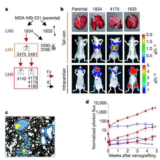
a , Flow chart of the selection of organ-specific metastatic subpopulations in vivo, indicating the organs from which these subpopulations were isolated. Each subsequent lung-metastatic generation is designated LM0, LM1 and LM2. The LM2 cells were further analysed for metastasis by either tail-vein (TV) or intracardiac (IC) xenografting. Metastatic propensities for all cell lines used in this study are listed in Supplementary Table 1. b, Representative lungs harvested at necropsy and BLI of the indicated cell lines are shown after tail-vein or intracardiac injection. The colour scale depicts the photon flux (photons per second) emitted from xenografted mice c, A representative image of haematoxylin staining of lung cryosections from mice injected with moderately metastatic 1834 cells showing an invading lesion (asterisk) and an embolus within the vascular space (arrowhead). Vascular walls are stained with the endothelial cell marker CD31. d, Parental cells (red) and 4175 (LM2) cells (blue) were tested for lung metastatic activity. Numbers of cells injected were as follows: diamonds, 2 × 10⁵; triangles, 2 × 10⁴; squares, 2 × 10³. Plots show a quantification of the luminescence signal as a function of time. Results are means ± s.e.m. for each cohort. Asterisks, P < 0.05 with a one-sided rank test, compared with mice injected with an equivalent number of parental cells.
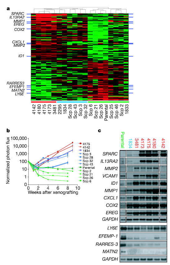
a, Comparison of gene expression profiles of LM2 populations with parental cells identifies 113 probe sets that are correlated with lung metastatic activity. This signature clusters populations selected in vivo and SCPs into groups that resemble the LM2 cell lines (red bar along the bottom), the parental MDA-MB-231 cell line (green bar) or an intermediate group (blue bar). b, LM2 populations 4175 and 4142 were assayed for lung metastatic activity as measured by BLI and were compared with parental populations and various SCPs. Plots show a quantification of the luminescence signal as a function of time. Results are means ± s.e.m. for each cohort. Colour-coding is as in a. c, Northern blot analysis of parental, LM0, LM1 and LM2 cell lines with a set of nine lung metastasis genes selected for functional validation, as well as four genes underexpressed in the lung-metastatic populations.
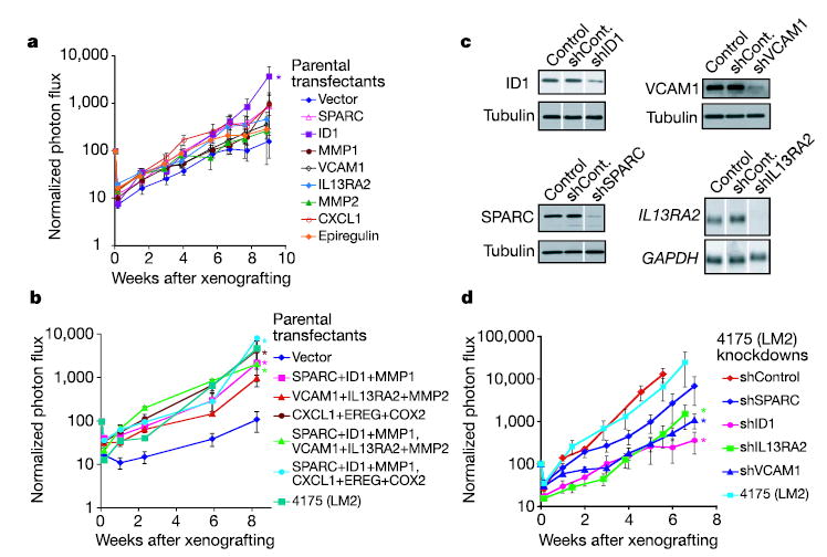
a, b, Retrovirus-mediated expression of selected genes from the lung metastasis signature in weakly metastatic parental MDA-MB-231 cells. Genes were tested individually (a) or in groups of three or six genes (b). c, Stable short hairpin (sh) RNAi constructs were introduced retrovirally into 4175 lung-metastatic cells, and their effectiveness at knocking down the expression of their intended target was validated at the protein level (ID1, VCAM1, SPARC) or at the mRNA level (IL13RA2). Controls were uninfected 4175 (LM2) cells, and shCont refers to 4175 cells transduced with a nonfunctional shRNAi. d, 4175 knockdown cell lines were xenografted through the tail vein to assess lung metastatic activity. One shRNAvector against ID1 that was ineffective at decreasing expression of this gene served as a negative control. Results are means ± s.e.m. for each cohort. Asterisks, P < 0.05 with a one-sided rank test.
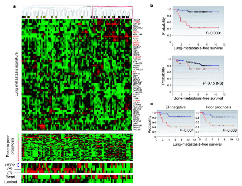
a, Hierarchical clustering of primary breast carcinomas from a cohort of 82 breast cancer patients was performed with the 54 lung metastasis signature genes. A dendrogram of the tumours is shown at the top, with tumours from patients who developed lung metastasis (black circles) or metastasis at non-pulmonary sites (yellow circles) denoted. A subcluster with a reproducibility index of 0.71 (dashed red box) groups tumours that tended to express the lung metastasis signature in a manner resembling the LM2 cell lines. The genes were also clustered; gene names are on the right. Functionally validated genes are in red. The Rosetta poor-prognosis signature is displayed with the genes underexpressed (green bar) and overexpressed (red bar) in poor-prognosis tumours indicated at the left. The expression of HER2, progesterone receptor (PR), oestrogen receptor (ER) and basal and luminal keratins is shown. Expression of the lung metastasis signature was confirmed in the independent Rosetta breast cancer cohort (Supplementary Fig. S6). b, Lung-metastasis-free survival and bone-metastasis-free survival for MSKCC patients who either expressed (red line) or did not express (blue line) the lung metastasis signature based on a classifier trained with the Rosetta cohort (Supplementary Fig. S7 and Supplementary Methods). The P value for each survival curve is shown. c, Lung-metastasis-free survival restricted to patients with ER-negative tumours or Rosetta-type poor-prognosis tumours.
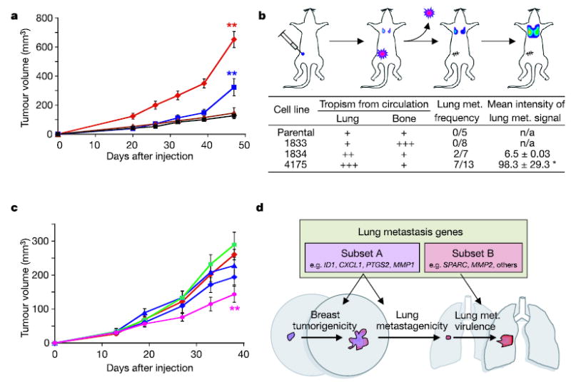
a, Representative MDA-MB-231 cell populations were injected into the mammary fat pad of immunodeficient mice and monitored for tumour growth. Red diamonds, 4175 cells (n = 9, where n is the number of mice in each cohort); blue squares, 1834 cells (n = 10); brown triangles, 1833 cells (n = 5); black squares, parental cells (n = 5). Each curve shows tumour volumes in cubic millimetres (means ± s.e.m.). b, As shown in the diagram, mice were inoculated with the indicated MDA-MB-231 cells into the mammary fat pad and tumours were removed after reaching a volume of 300 mm3. Lung metastasis was monitored with BLI, and normalized photon flux was measured 2 weeks after removal of the primary tumour. Asterisk, a mouse in the 4175 cohort with an unusually high normalized photon flux of 36,400 was excluded. c, Growth in mammary fat pad of highly lung- metastatic 4175 (LM2) cells after stable shRNA knockdown of the following gene products: red diamonds, shControl; blue triangles, shVCAM; green squares, shIL13RA2; blue diamonds, shSPARC; pink circles, shID1. shControl refers to a cell line transduced with a short hairpin construct that did not result in effective knockdown of its target gene. Two asterisks, P < 0.01 by a one-sided rank test. Each curve shows tumour volumes in cubic millimetres (means ± s.e.m.). d, A model of two classes of genes contained within the lung metastasis signature. The first class (subset A) confers both breast tumorigenicity and basal lung metastagenicity. Examples may include ID1, CXCL1, PTGS2 and MMP1. The second class (subset B) confers functions specific to the lung microenvironment, facilitating lung metastatic virulence. Examples may include SPARC and MMP2.
References
-
- Chambers AF, Groom AC, MacDonald IC. Dissemination and growth of cancer cells in metastatic sites. Nature Rev Cancer. 2002;2:563–572. - PubMed
-
- Fidler IJ. The pathogenesis of cancer metastasis: the ‘seed and soil’ hypothesis revisited. Nature Rev Cancer. 2003;3:453–458. - PubMed
-
- Yokota J. Tumor progression and metastasis. Carcinogenesis. 2000;21:497–503. - PubMed
-
- Kang Y, et al. A multigenic program mediating breast cancer metastasis to bone. Cancer Cell. 2003;3:537–549. - PubMed
-
- Clark EA, Golub TR, Lander ES, Hynes RO. Genomic analysis of metastasis reveals an essential role for RhoC. Nature. 2000;406:532–535. - PubMed
Publication types
MeSH terms
Substances
Grants and funding
LinkOut - more resources
Full Text Sources
Other Literature Sources
Medical
Molecular Biology Databases
Research Materials