. 2005 Nov 15;392(Pt 1):145-52.
doi: 10.1042/BJ20050412.

Heat-shock cognate 70 is required for the activation of heat-shock factor 1 in mammalian cells

Affiliations

Heat-shock cognate 70 is required for the activation of heat-shock factor 1 in mammalian cells

Sang-Gun Ahn et al. Biochem J. .

Abstract

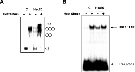
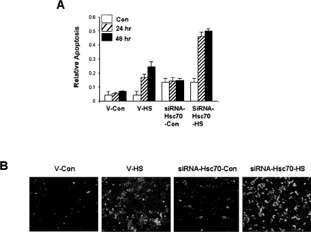
HSF1 (heat-shock factor 1) plays an essential role in mediating the appropriate cellular response to diverse forms of physiological stresses. However, it is not clear how HSF1 is regulated by interacting proteins under normal and stressful conditions. In the present study, Hsc70 (heat-shock cognate 70) was identified as a HSF1-interacting protein using the TAP (tandem affinity purification) system and MS. HSF1 can interact with Hsc70 in vivo and directly in vitro. Interestingly, Hsc70 is required for the regulation of HSF1 during heat stress and subsequent target gene expression in mammalian cells. Moreover, cells transfected with siRNAs (small interfering RNAs) targeted to Hsc70 showed greatly decreased HSF1 activation with expression of HSF1 target genes being dramatically reduced. Finally, loss of Hsc70 expression in cells resulted in an increase in stress-induced apoptosis. These results indicate that Hsc70 is a necessary and critical regulator of HSF1 activities.


Figures

Figure 1
Figure 1. Identification of HSF1-interacting proteins
(A) HEK-293 cells were transfected with either pCDNA3.1-TAP (C) or pCDNA3.1-HSF1-TAP (HSF1) expression vector. At 48 h after transfection, TAP-tagged HSF1 and its associated proteins were purified using the TAP system. Co-purified proteins were separated by SDS/PAGE, and the gel was stained with Coomassie Blue. Two bands excised for MALDI–TOF MS analysis are marked with an asterisk (*). Molecular-mass sizes are indicated in kDa. (B) MALDI–TOF MS of the tryptic digests of Hsc70. Shown in bold are peptide masses unique to p71 (upper band in A) that were sequenced by MALDI–PSD (labelled MS/MS). (C) HEK-293 cells were transfected with TAP-tagged HSF1-(1–290), TAP-tagged HSF1 (FL) expression vectors or empty vector control (C). At 48 h after transfection, cell extracts were subjected to immunoprecipitation using the TAP system, and endogenous Hsc70 was detected by Western blot (WB) analysis using anti-Hsc70 antibodies. (D) Purified GST-tagged bacterial recombinant HSF1-(1–290) or HSF1 (FL) (1 μg) were incubated with equimolar His6-tagged bacterial recombinant Hsc70. HSF1 was pulled-down using glutathione–agarose beads. Co-precipitated proteins were resolved by SDS/PAGE and detected by Western blot (WB) analysis using anti-His antibodies. C, empty vector control. (E) HEK-293 cells were transfected with TAP-tagged HSF1 (FL) expression vector. At 48 h after transfection, cells were untreated (−) or heat-shocked at 42 °C (+) for 1 h. Cell extracts were subjected to immunoprecipitation using the TAP system, and endogenous Hsc70 was detected by Western blot analysis using anti-Hsc70 antibodies.
Figure 2
Figure 2. Hsc70 interacts with HSF1 through its C-terminal region
(A) Schematic diagram of Hsc70 domain organization. Hsc70 is composed of an N-terminal ATPase domain, a central substrate-binding domain and a C-terminal domain of unknown function. The substrate-binding domain contains a coiled-coil domain that is likely to facilitate protein–protein interactions. Domain boundaries were obtained from the SMART and COIL programs. (B) HEK-293 cells were transfected with the indicated TAP-tagged expression vectors or empty vector control (C). At 48 h after transfection, TAP-tagged Hsc70 was purified, and co-precipitated endogenous HSF1 was analysed by Western blot analysis using anti-HSF1 antibodies (upper panel). The expression level of proteins in the transfected cells was monitored by Western blot analysis using anti-Hsc70 antibodies (lower panel). Molecular-mass sizes are given in kDa.
Figure 3
Figure 3. Hsc70 affects HSF1-trimerization and DNA-binding activities
HEK-293 cells were transfected with either pCDNA3.1 (C) or pCDNA3.1-FLAG-Hsc70 mammalian expression vector. At 48 h after transfection, cells were untreated (−) or heat shocked at 42 °C (+) for 1 h. (A) Whole-cell extracts (20 μg) were subjected to in vitro cross-linking experiment with 2 mM EGS. HSF1 was detected by Western blot analysis using anti-HSF1 antibodies. The positions of HSF1 monomers, dimers, trimers and putative hexamers (6×) are shown on the right. (B) Nuclear extracts were prepared, and HSF1 DNA-binding activity was measured by EMSA using 32P-labelled HSF1 DNA-binding fragments known as HSEs. The position of the HSF1–HSE complex and the free HSE DNA fragments are shown on the right. C, empty vector control.
Figure 4
Figure 4. HSF1 and Hsc70 are co-localized in the nucleus after heat shock
(A) WT and (B) hsf1−/− mEF cells were heat-shocked at 42 °C for 1 h. Cytosolic (C) and nuclear (N) fractions were prepared and resolved by SDS/PAGE. HSF1, Hsc70, actin and c-fos were detected by Western blot analysis. (C) hsf1−/− mEF cells were co-transfected with pEGFP-HSF1 and pCDNA3.1-FLAG-Hsc70 mammalian expression vectors. At 48 h after transfection, cells were untreated (Control) or heat-shocked at 42 °C for 1 h. Cells were fixed and immunostained using anti-FLAG antibodies. Nuclei were stained with DAPI (4,6-diamidino-2-phenylindole).
Figure 5
Figure 5. Depletion of endogenous Hsc70 by RNAi inhibits heat-stress-mediated HSF1 activation
HEK-293 cells were transiently transfected with a pSuppressorNeo (C) or pSuppressorNeo-Hsc70 RNAi expression vector. (A) At 48 h after transfection, total cell extracts were prepared, and Hsc70 and actin protein levels were detected by Western blot analysis. (B) At 48 h after transfection, cells were untreated (−) or heat-shocked at 42 °C (+) for 1 h. Nuclear extracts were prepared, and HSF1 DNA-binding activity was measured by EMSA using 32P-labelled HSF1 DNA-binding fragments known as HSEs. The position of the HSF1–HSE complex and the free HSE DNA fragments are shown on the right. (C) At 48 h after transfection, cells were untreated (−) or heat-shocked at 42 °C (+) for 1 h, followed by recovery at 37 °C for 24 or 48 h. Total cell extracts were prepared, and Hsp70, Hsp27, HSF1 and actin protein levels were detected by Western blot analysis.
Figure 6
Figure 6. Depletion of endogenous Hsc70 by RNAi leads to heat-stress-induced apoptosis
HEK-293 cells were transiently transfected with a pSuppressorNeo (V) or pSuppressorNeo-Hsc70 RNAi expression vector. (A) At 48 h after transfection, cells were untreated (Con) or heat-shocked at 44 °C (HS) for 1 h, followed by recovery at 37 °C for 24 or 48 h. Relative cell death was measured as a ‘relative apoptosis’ index, see the Materials and methods section for details. (B) At 48 h after transfection, cells were untreated (Con) or heat-shocked at 44 °C (HS) for 1 h and then cultured for a further 24 h at 37 °C. Apoptosis was detected by annexin V–FLUOS and propidium iodide staining.

Similar articles

Cited by

References

    1. Parsell D. A., Lindquist S. The function of heat-shock proteins in stress tolerance: degradation and reactivation of damaged proteins. Annu. Rev. Genet. 1993;27:437–496. - PubMed
    1. Zugel U., Kaufmann S. H. Role of heat shock proteins in protection from and pathogenesis of infectious diseases. Clin. Microbiol. Rev. 1999;12:19–39. - PMC - PubMed
    1. Morimoto R. I., Tissieres A., Georgopoulos C. Cold Spring Harbor Monograph Series, vol. 26. Cold Spring Harbor: Cold Spring Harbor Laboratory Press; 1994. The biology of the heat shock proteins and molecular chaperones; p. 610.
    1. Fink A. L. Chaperone-mediated protein folding. Physiol. Rev. 1999;79:425–449. - PubMed
    1. Kaufman R. J. Molecular chaperones and the heat shock response. Sponsored by Cold Spring Harbor Laboratory, 6–10 May 1998. Biochim. Biophys. Acta. 1999;1423:R13–R27. - PubMed