Adenomyoepithelial tumours and myoepithelial carcinomas of the breast--a spectrum of monophasic and biphasic tumours dominated by immature myoepithelial cells
- PMID: 16050957
- PMCID: PMC1187882
- DOI: 10.1186/1471-2407-5-92
Adenomyoepithelial tumours and myoepithelial carcinomas of the breast--a spectrum of monophasic and biphasic tumours dominated by immature myoepithelial cells
Abstract
Background: Adenomyoepithelial tumours and myoepithelial carcinomas of the breast are primarily defined by the presence of neoplastic cells with a myoepithelial immunophenotype. Current classification schemes are based on purely descriptive features and an assessment of individual prognosis is still problematic.
Methods: A series of 27 adenomyoepithelial tumours of the breast was analysed immunohistochemically with antibodies directed against various cytokeratins, p63, smooth muscle alpha-actin (SMA) and vimentin. Additionally, double immunofluorescence and comparative genomic hybridisation (CGH) was performed.
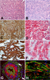
Results: Immunohistochemically, all the tumours showed a constant expression of high molecular weight cytokeratins (Ck) Ck5 and Ck14, p63, SMA and vimentin. With exception of one case diagnosed as myoepithelial carcinoma, all tested tumours expressed low molecular weight cytokeratin Ck18 in variable proportions of cells. Even in monophasic tumours lacking obvious glandular differentiation in conventional staining, a number of neoplastic cells still expressed those cytokeratins. Double immunofluorescence revealed tumour cells exclusively staining for Ck5/Ck14 in the presence of other cell populations that co-expressed high molecular weight Ck5/Ck14 as well as either low molecular weight Ck8/18 or SMA. Based on morphology, we assigned the series to three categories, benign, borderline and malignant. This classification was supported by a stepwise increase in cytogenetic alterations on CGH.
Conclusion: Adenomyoepithelial tumours comprise a spectrum of neoplasms consisting of an admixture of glandular and myoepithelial differentiation patterns. As a key component SMA-positive cells co-expressing cytokeratins could be identified. Although categorisation of adenomyoepithelial tumours in benign, borderline and malignant was supported by results of CGH, any assessment of prognosis requires to be firmly based on morphological grounds. At present it is not yet clear, if and to what extent proposed Ck5-positive progenitor cells contribute to the immunohistochemical and morphological heterogeneity of these neoplasms of the breast.
Figures


Similar articles
-
Morphological and immunophenotypic analysis of breast carcinomas with basal and myoepithelial differentiation.J Pathol. 2006 Mar;208(4):495-506. doi: 10.1002/path.1916. J Pathol. 2006. PMID: 16429394
-
Cell blocks allow reliable evaluation of expression of basal (CK5/6) and luminal (CK8/18) cytokeratins and smooth muscle actin (SMA) in breast carcinoma.Cytopathology. 2010 Aug;21(4):259-66. doi: 10.1111/j.1365-2303.2009.00713.x. Epub 2009 Oct 15. Cytopathology. 2010. PMID: 19843143
-
The dog as a natural animal model for study of the mammary myoepithelial basal cell lineage and its role in mammary carcinogenesis.J Comp Pathol. 2014 Aug-Oct;151(2-3):166-80. doi: 10.1016/j.jcpa.2014.04.013. Epub 2014 Jun 27. J Comp Pathol. 2014. PMID: 24975897
-
Myoepithelial carcinoma of intraoral minor salivary glands: a clinicopathological study of 7 cases and review of the literature.Oral Surg Oral Med Oral Pathol Oral Radiol Endod. 2010 Jul;110(1):85-93. doi: 10.1016/j.tripleo.2010.02.023. Epub 2010 May 21. Oral Surg Oral Med Oral Pathol Oral Radiol Endod. 2010. PMID: 20488733 Review.
-
The usefulness of p63 as a marker of breast myoepithelial cells.In Vivo. 2003 Nov-Dec;17(6):573-6. In Vivo. 2003. PMID: 14758723 Review.
Cited by
-
[Salivary gland-like tumors of the breast].Pathologe. 2006 Sep;27(5):363-72. doi: 10.1007/s00292-006-0851-0. Pathologe. 2006. PMID: 16896677 German.
-
Immunohistochemical expression of myoepithelial markers in adenomyoepithelioma of the breast: a unique paradoxical staining pattern of high-molecular weight cytokeratins.Virchows Arch. 2015 Feb;466(2):191-8. doi: 10.1007/s00428-014-1687-2. Epub 2014 Dec 6. Virchows Arch. 2015. PMID: 25479938
-
The prognostic significance of p63 and Ki-67 expression in myoepithelial carcinoma.Head Neck Oncol. 2012 Mar 27;4:9. doi: 10.1186/1758-3284-4-9. Head Neck Oncol. 2012. PMID: 22452794 Free PMC article.
-
Malignant adenomyoepithelioma combined with adenoid cystic carcinoma of the breast: a case report and literature review.Diagn Pathol. 2014 Jul 23;9:148. doi: 10.1186/1746-1596-9-148. Diagn Pathol. 2014. PMID: 25056281 Free PMC article. Review.
-
A block in lineage differentiation of immortal human mammary stem / progenitor cells by ectopically-expressed oncogenes.J Carcinog. 2011;10:39. doi: 10.4103/1477-3163.91415. Epub 2011 Dec 31. J Carcinog. 2011. PMID: 22279424 Free PMC article.
References
-
- Tavassoli FA. Pathology of the breast. Norwalk, Conneticut, USA: Appleton & Lange; 1992. Myoepithelial lesions of the breast; pp. 593–609.
-
- Tavassoli FA. Myoepithelial lesions of the breast. Myoepitheliosis, adenomyoepithelioma, and myoepithelial carcinoma. Am J Surg Pathol. 1991;15:554–68. - PubMed
-
- Tavassoli FA. Pathology and Genetics of Tumours of the Breast and Female Genital Organs. World Health Organisation. IARC; 2003.
-
- Bult P, Verwiel JM, Wobbes T, Kooy-Smits MM, Biert J, Holland R. Malignant adenomyoepithelioma of the breast with metastasis in the thyroid gland 12 years after excision of the primary tumor. Case report and review of the literature. Virchows Arch. 2000;436:158–166. - PubMed
-
- Jones C, Foschini MP, Chaggar R, Lu YJ, Wells D, Shipley JM, Eusebi V, Lakhani SR. Comparative genomic hybridization analysis of myoepithelial carcinoma of the breast. Lab Invest. 2000;80:831–836. - PubMed
Publication types
MeSH terms
Substances
LinkOut - more resources
Full Text Sources
Medical
Research Materials

