. 2005 Sep 30;340(2):209-23.
doi: 10.1016/j.virol.2005.06.035.

Mutagenesis of the murine hepatitis virus nsp1-coding region identifies residues important for protein processing, viral RNA synthesis, and viral replication

Affiliations

Mutagenesis of the murine hepatitis virus nsp1-coding region identifies residues important for protein processing, viral RNA synthesis, and viral replication

Sarah M Brockway et al. Virology. .

Abstract

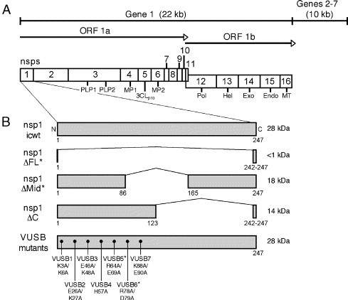
Despite ongoing research investigating mechanisms of coronavirus replication, functions of many viral nonstructural proteins (nsps) remain unknown. In the current study, a reverse genetic approach was used to define the role of the 28-kDa amino-terminal product (nsp1) of the gene 1 polyprotein during replication of the coronavirus murine hepatitis virus (MHV) in cell culture. To determine whether nsp1 is required for MHV replication and to identify residues critical for protein function, mutant viruses that contained deletions or point mutations within the nsp1-coding region were generated and assayed for defects in viral replication, viral protein expression, protein localization, and RNA synthesis. The results demonstrated that the carboxy-terminal half of nsp1 (residues K(124) through L(241)) was dispensable for virus replication in culture but was required for efficient proteolytic cleavage of nsp1 from the gene 1 polyprotein and for optimal viral replication. Furthermore, whereas deletion of nsp1 residues amino-terminal to K(124) failed to produce infectious virus, point mutagenesis of the nsp1 amino-terminus allowed recovery of several mutants with altered replication and RNA synthesis. This study identifies nsp1 residues important for protein processing, viral RNA synthesis, and viral replication.


Figures

Fig. 1
Fig. 1
Schematics of the MHV gene 1 polyproteins and nsp1 mutants. (A) Organization of the MHV gene 1 polyprotein. The 32-kb MHV genome is shown as a line, and the locations of gene 1 (22 kb) and genes 2–7 (10 kb) are indicated. Gene 1 is composed of two open reading frames (ORF1a and ORF1b). The ORF1a–ORF1b fusion polyprotein is illustrated with mature nonstructural protein (nsps) represented as numbered boxes. The gray box represents the amino-terminal cleavage product (nsp1). Nsps with confirmed or predicted functions include: two papain-like proteinases (PLP1 and PLP2 within nsp3), the 3C-like proteinase (3CLpro; nsp5), two trans-membrane proteins (MP1 and MP2; nsp4 and nsp6, respectively), the RNA-dependent RNA polymerase (Pol; nsp12), the RNA helicase (Hel; nsp13), the 3′-to-5′ exonuclease (Exo; nsp14), the endoribonuclease (Endo; nsp15), and the RNA methyltransferase (MT; nsp16). (B) Nsp1 mutant proteins. The schematics illustrate the engineered deletions and point mutations within nsp1. Nsp1 amino acid numbers are listed below each protein, and the predicted protein size (in kilodaltons) is listed to the right of each. The amino-terminal charge-to-alanine mutations for each VUSB mutants are listed below the bottom nsp1 protein. The asterisks (*) indicate mutants that did not establish productive infections as determined by lack of recovered virus from electroporated cells.
Fig. 2
Fig. 2
Single-cycle replication of nsp1 mutant viruses. (A) Replication kinetics. Wild-type (icwt) or nsp1 mutant viruses were used to infect DBT-9 cells at an MOI of 5. Cells were rinsed three times with PBS, incubated under medium at 37 °C, and samples of medium were obtained at the indicated times post-infection. Viral titers in each sample were determined using plaque assays on DBT-9 cells at 37 °C. The graph shows the results of a representative experiment. Values are the averages obtained from duplicate media samples. (B) Nsp1 mutant viral yield. To determine viral yield, viral titer at 1 h p.i. was subtracted from peak titer for each virus. Bars represent average viral yield calculated from the experiments (n = 3). Lines represent standard error from the experiments. Statistical analysis software was used to determine P values using a two-sample t test. Asterisks (*) indicate P values ≤ 0.05. (C) Relative plaque size of icwt, VUSB1, and VUSB4 (15 h p.i.). Images were obtained at the same resolution (10×) on a Nikon Eclipse TE2000-E microscope. White circles were drawn to facilitate visualization of the plaque boundary.
Fig. 3
Fig. 3
Nsp1 mutant viral protein expression and processing. Cytoplasmic lysates were generated from radiolabeled DBT-9 cells that were either mock-infected (m) or infected with icwt or nsp1 mutant viruses. Labeled proteins were immunoprecipitated from cytoplasmic lysates with the indicated polyclonal antisera. Proteins were resolved by SDS-PAGE in 5–18% polyacrylamide gradient gels and visualized following fluorography. Images were obtained following 4-day film exposure. Bands corresponding to unique or predicted proteins are indicated on the right of the fluorograms, and molecular weight standards (in kilodaltons) are shown on the left. The MHV structural proteins are designated as follows: spike (S), nucleocapsid (N), and membrane (M). The antisera used for immunoprecipitation are indicated: (A) α-nsp1; (B) α-nsp2; (C) α-nsp8; and (D) α-MHV.
Fig. 4
Fig. 4
Pulse-chase translation in nsp1ΔC virus-infected cells. Proteins in infected DBT-9 cells were radiolabeled for 30 min with [35S]Met/Cys at 6 h p.i. and then incubated in medium containing cyclohexamide for 15 to 360 min as described in Materials and methods. Cells were lysed at the indicated times (min) post-chase (p.c.), and cytoplasmic lysates were generated for immunoprecipitation studies using α-nsp1 and α-nsp2. Proteins were analyzed as in Fig. 3. The identities of proteins are indicated to the right of the fluorograms. The number of days the gels were exposed to film (d exp) to generate the image is listed next to the antisera used for immunoprecipitation. Pulse-chase translation in cells infected with (A) icwt or (B) nsp1ΔC virus.
Fig. 5
Fig. 5
Intracellular localization of mutant nsp1 proteins. DBT-9 cells grown on glass coverslips were infected with icwt or nsp1 mutant viruses for 7 h, fixed and permeablized with 100% methanol, and incubated with antibodies against nsp1 (red), nsp2 (green), and N (purple). Cells were imaged using a Zeiss LSM 510 confocal microscope at 546 nm (red), 488 nm (green), and 633 nm (purple). Images are single confocal slices obtained using a 40× objective. Co-localization of green and purple for VUSB1 is shown in the merged image as light green pixels. Co-localization of all three colors is shown in the merged images as white pixels. Multi-nucleated cells are a cytopathic effect of MHV replication.
Fig. 6
Fig. 6
Nsp1 mutant viral RNA levels. (A) Nsp1 mutant viral RNA levels represented as percent of icwt. DBT-9 cells were infected at an MOI of 5 with icwt or nsp1 mutant viruses. Actinomycin D was added to a final concentration of 20 μg/ml 30 min prior to the addition of [3H]uridine. Viral RNA was radiolabeled from 5–9 h p.i. and then precipitated from equal volumes of cytoplasmic lysates in replicate using trichloracetic acid. To quantitate [3H]uridine incorporation as counts per minute (CPM), liquid scintillation was used. For each experiment (n = 5), labeled viral RNA levels (CPM) for icwt were set to 100%, and nsp1 mutant viral RNA was calculated as a percentage of the icwt value. The bars represent the average percent viral RNA from all experiments, and lines indicate standard error. Statistical analysis software was used to determine P values using a one-sample t test. Asterisks (*) indicate P values ≤ 0.05. (B) Gel analysis of viral RNA. DBT-9 cells were mock-infected or infected with icwt or the indicated nsp1 mutants. RNA was labeled as in panel (A) above, and cells were lysed using Trizol. RNA was isolated from cell lysates and normalized so as to electrophorese the same amount of radiolabeled viral RNA for each mutant (approximately 400,000 CPM from a maximum of 106 cells). For the mock-infected control (m), RNA from 106 cells was used. The RNA was separated in an 0.8% formaldehyde/agarose gels and individual RNA species were visualized following fluorography. The image is from a 2-day exposure of the gel to film. Individual viral RNA species are numbered to the right of the fluorogram (RNA 1 = genome and RNA 2–7 = subgenomic RNAs). The asterisk (*) indicates a unique RNA band detected in nsp1ΔC virus-infected cells.
Fig. 7
Fig. 7
Model of icwt and nsp1ΔC virus protein processing. Shown are schematics of wild-type and nsp1ΔC virus amino-terminal gene 1 nsps (boxes). PLP1 and PLP2 are shown as gray boxes within nsp3. The individual cleavage sites are labeled, and the order of processing is indicated above the closed arrowheads. Lines below the protein schematics illustrate the size of mature proteins following cleavage at individual sites. (A) Processing of wild-type nsp1, nsp2, and nsp3. During infection, nsp1 (28 kDa) is cleaved rapidly at cleavage site 1 (CS1) as the polyprotein is translated (indicated by forward slashes). Next, cleavage site 3 (CS3) is processed to yield a 275-kDa nsp2–nsp3 precursor. Finally, cleavage site 2 (CS2) is cleaved to liberate nsp2 (65 kDa) and nsp3 (210 kDa). (B) Wild-type pattern of processing for nsp1ΔC mutant. The order of cleavage for nsp1ΔC mutant polyprotein is identical to that of icwt. Nsp1ΔC protein (ΔC; 14 kDa) is liberated by CS1 cleavage. Next, CS3 is processed to yield a 275-kDa nsp2–nsp3 precursor. Finally, CS2 is cleaved to liberate nsp2 (65 kDa) and nsp3 (210 kDa). (C) Alternative pattern of processing for nsp1ΔC mutant. If CS1 is not initially cleaved, a 290-kDa nsp1ΔC–nsp2–nsp3 precursor is made following CS3 processing. This precursor is then cleaved at CS2 to yield an 80-kDa protein (nsp1ΔC–nsp2) and nsp3.

Similar articles

Cited by

References

    1. Appel N., Herian U., Bartenschlager R. Efficient rescue of hepatitis C virus RNA replication by trans-complementation with nonstructural protein 5A. J. Virol. 2005;79:896–909. - PMC - PubMed
    1. Baker S.C., Shieh C.-K., Soe L.H., Chang M.-F., Vannier D.M., Lai M.M.C. Identification of a domain required for autoproteolytic cleavage of murine coronavirus gene A polyprotein. J. Virol. 1989;63:3693–3699. - PMC - PubMed
    1. Baker S.C., Yokomori K., Dong S., Carlisle R., Gorbalenya A.E., Koonin E.V., Lai M.M. Identification of the catalytic sites of a papain-like cysteine proteinase of murine coronavirus. J. Virol. 1993;67:6056–6063. - PMC - PubMed
    1. Bhardwaj K., Guarino L., Kao C.C. The severe acute respiratory syndrome coronavirus Nsp15 protein is an endoribonuclease that prefers manganese as a cofactor. J. Virol. 2004;78:12218–12224. - PMC - PubMed
    1. Bonilla P.J., Gorbalenya A.E., Weiss S.R. Mouse hepatitis virus strain A59 RNA polymerase gene ORF 1a: heterogeneity among MHV strains. Virology. 1994;198:736–740. - PMC - PubMed

Publication types

LinkOut - more resources