. 2005 Sep 23;280(38):32655-61.
doi: 10.1074/jbc.M504778200. Epub 2005 Jul 28.

Cartilage oligomeric matrix protein/thrombospondin 5 supports chondrocyte attachment through interaction with integrins

Affiliations

Cartilage oligomeric matrix protein/thrombospondin 5 supports chondrocyte attachment through interaction with integrins

Faye Hui Chen et al. J Biol Chem. .

Abstract

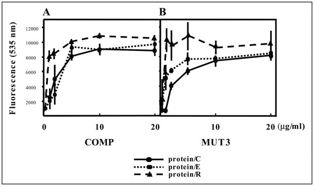
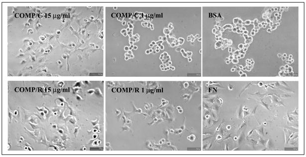
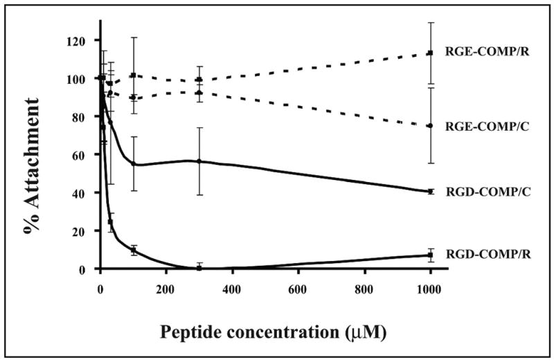
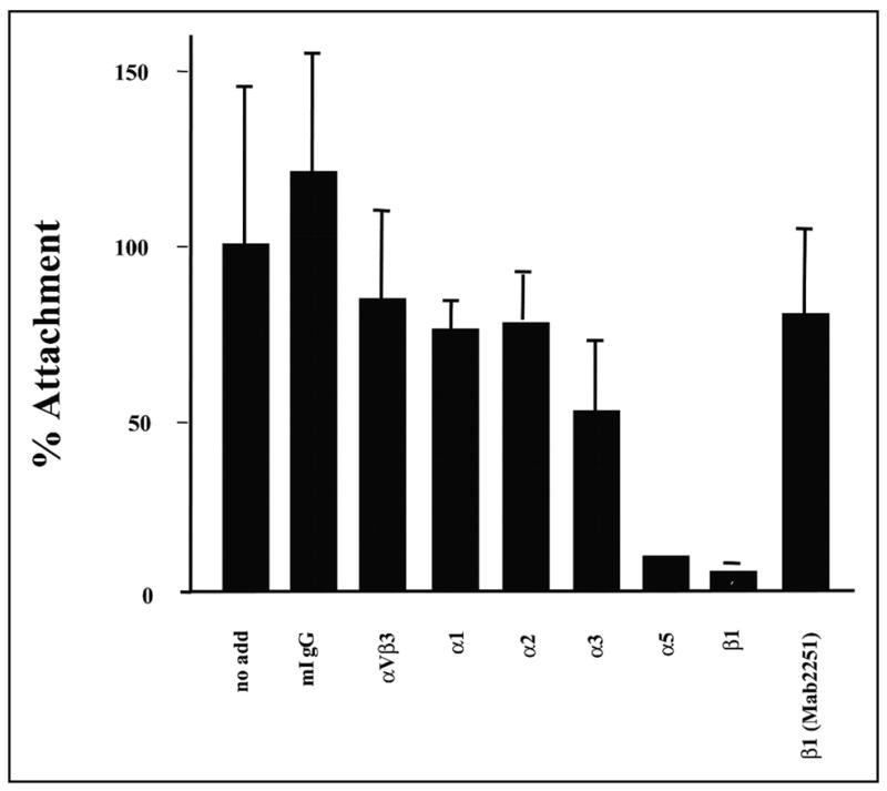
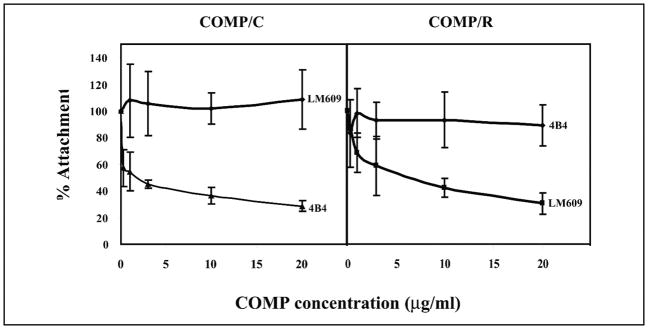
Cartilage oligomeric matrix protein/thrombospondin 5 (COMP/TSP5) is a major component of the extracellular matrix of the musculoskeletal system. Although COMP/TSP5 abnormalities are associated with several pathological conditions, its normal function remains unclear. This study was undertaken to delineate the function(s) of COMP/TSP5 in cartilage, especially regarding its interaction with chondrocytes. We show that COMP/TSP5 can support chondrocyte attachment and that the RGD sequence in COMP/TSP5 and the integrin receptors alpha5beta1 and alphaVbeta3 on the chondrocytes are involved in mediating this attachment. The interactions of COMP/TSP5 with the integrins are dependent on COMP/TSP5 conformation. Chondrocyte attachment to COMP/TSP5 in the calcium-replete conformation was inhibited by function-blocking integrin alpha5 and beta1 antibodies, suggesting the involvement of the alpha5beta1 integrin. Under this condition, a function-blocking antibody against alphaVbeta3 did not have any effect on cell attachment. On the other hand, chondrocyte attachment to reduced COMP/TSP5 was instead sensitive to alphaVbeta3 function-blocking antibodies, suggesting that COMP/TSP5 mediates attachment through chondrocyte alphaVbeta3 integrin under this condition. Cell attachment to reduced COMP/TSP5 was not inhibited by beta1 antibodies. These data indicate that COMP/TSP5 in different conformations can utilize different integrin receptors. These results are the first to demonstrate that COMP/TSP5 can mediate chondrocyte attachment through interactions with integrins. Through these interactions, COMP/TSP5 may be able to regulate cellular activities and respond to environment in the surrounding cartilage matrix.


Figures

FIGURE 1
FIGURE 1. Attachment of chondrocyte-derived cell line TC1a cultured in monolayer to COMP/TSP5 (A) and MUT3 (B)
COMP/TSP5 or MUT3 in the presence of 1 mM CaCl2 (protein/C) or 5 mM EDTA (protein/E) or coated in the presence of 5 mM EDTA followed by reduction (protein/R) were diluted to the final concentrations as indicated and coated onto 96-well plates. Attachment assays were carried out as described under ”Materials and Methods“ with TC1a cells grown in a monolayer. Results are shown as mean ± S.D. of quadruplicate experiments.
FIGURE 2
FIGURE 2. Morphology of TC1a attached to COMP/TSP5
TC1a cells were allowed to attach to COMP/TSP5 coated at the indicated concentrations in the presence of 1 mM CaCl2 (COMP/C) or 5 mM EDTA followed by reduction (COMP/R). FN coated at 10 μg/ml (FN) or heat-inactivated bovine serum albumin (BSA) coated at the same concentration was used as control. Attachment assays were carried out as described under ”Materials and Methods.“ At the end of the 2-h incubation, phase-contrast pictures were taken.
FIGURE 3
FIGURE 3. Effect of cycloheximide on attachment of chondrocyte-derived cell line TC1a
Attachment assays were carried out as described in the Fig. 1 legend with COMP/TSP5 coated in the presence of 1 mM CaCl2. TC1a cells were cultured in serum free Dulbecco’s modified Eagle’s medium containing 20 μg/ml cycloheximide 2 h before the attachment assay, and cycloheximide was included in all buffers for the attachment assay. Results are shown as mean ± S.D. of six data points.
FIGURE 4
FIGURE 4. Effect of RGD peptide on attachment of chondrocyte-derived cell line TC1a to COMP/TSP5
Attachment assays were carried out as described in the Fig. 1 legend with COMP/TSP5 coated in the presence of CaCl2 (COMP/C) or EDTA followed by reduction (COMP/R). Primary chondrocytes were used for this attachment assay. Cells were incubated with either the GRGDSP (RGD) or GRGESP (RGE) peptide for 30 min at the indicated concentrations before being added to the COMP/TSP5-coated wells.
FIGURE 5
FIGURE 5. Integrin expression by the chondrocytes
Primary chondrocyte cell surface proteins were labeled with biotin. The cells were lysed, and integrins were immunoprecipitated using specific integrin antibodies as indicated. The immunoprecipitated integrins were separated by SDS-PAGE followed by transfer to a piece of nitrocellulose membrane. The biotin-labeled integrins were visualized by horseradish peroxidase-conjugated streptavidin incubation followed by enhanced chemiluminescence detection. mIgG, mouse IgG.
FIGURE 6
FIGURE 6. Attachment of chondrocytes to COMP/TSP5/C in the presence of integrin antibodies
Attachment assays were carried out basically as described in the Fig. 1 legend. COMP/TSP5 in the presence of CaCl2 was coated on the wells. The primary chondrocytes used for this attachment assay were incubated with the various antibodies for 30 min before addition to the wells. Results are shown as mean ± S.D. of two triplicate experiments. The monoclonal antibodies used in this experiment and their final concentrations are as follows: mouse IgG (mIgG), 100 μg/ml; anti-α1, 6.3 μg/ml; anti-α2, 6 μg/ml; anti-α5,16.6 μg/ml; anti-α6, 16.6 μg/ml; anti-β1 (P4C10), 16.6 μg/ml; and anti-β1 (B3B11), 16.6 μg/ml.
FIGURE 7
FIGURE 7. Effect of COMP/TSP5 conformation on integrin usage by chondrocytes for attachment
Attachment assays were carried out basically as described in the Fig. 1 legend. COMP/TSP5 was either coated in the presence of CaCl2 (COMP/C) or coated in the presence of EDTA followed by reduction (COMP/R) before the attachment assays. Chondrocytes were incubated with the different antibodies at the indicated concentrations for 30 min before addition to the wells. Shown here are the results using TC1a cells from a monolayer culture.

References

    1. DiCesare PE, Fang C, Leslie MP, Tulli H, Perris R, Carlson CS. J Orthop Res. 2000;18:713–720. - PubMed
    1. DiCesare PE, Carlson CS, Stollerman ES, Chen FS, Leslie M, Perris R. FEBS Lett. 1997;412:249–252. - PubMed
    1. DiCesare P, Hauser N, Lehman D, Pasumarti S, Paulsson M. FEBS Lett. 1994;354:237–240. - PubMed
    1. Hedbom E, Antonsson P, Hjerpe A, Aeschlimann D, Paulsson M, Rosa-Pimentel E, Sommarin Y, Wendel M, Oldberg A, Heinegard D. J Biol Chem. 1992;267:6132–6136. - PubMed
    1. Hecht JT, Deere M, Putnam E, Cole W, Vertel B, Chen H, Lawler J. Matrix Biol. 1998;17:269–278. - PubMed

Publication types

MeSH terms