Comparative Study
. 2005 Aug 9;102(32):11539-44.
doi: 10.1073/pnas.0504926102. Epub 2005 Jul 28.

Organically modified silica nanoparticles: a nonviral vector for in vivo gene delivery and expression in the brain

Affiliations
Comparative Study

Organically modified silica nanoparticles: a nonviral vector for in vivo gene delivery and expression in the brain

Dhruba J Bharali et al. Proc Natl Acad Sci U S A. .

Abstract

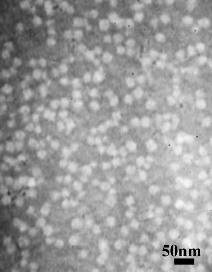
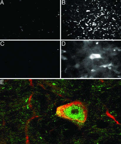
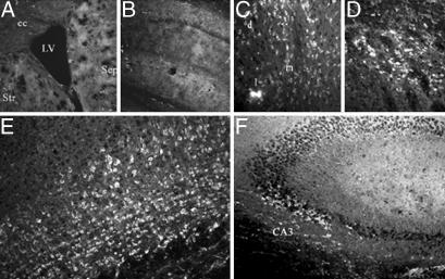
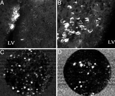
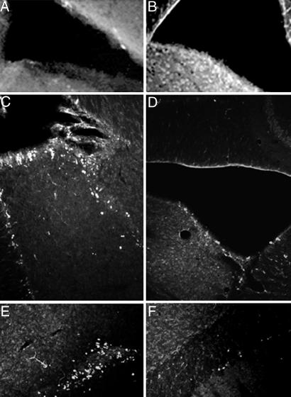
This article reports on the application of organically modified silica (ORMOSIL) nanoparticles as a nonviral vector for efficient in vivo gene delivery. Highly monodispersed, stable aqueous suspension of nanoparticles, surface-functionalized with amino groups for binding of DNA, were prepared and characterized. Stereotaxic injections of nanoparticles, complexed with plasmid DNA encoding for EGFP, into the mouse ventral midbrain and into lateral ventricle, allowed us to fluorescently visualize the extensive transfection of neuronal-like cells in substantia nigra and areas surrounding the lateral ventricle. No ORMOSIL-based toxicity was observed 4 weeks after transfection. The efficiency of transfection equaled or exceeded that obtained in studies using a viral vector. An in vivo optical imaging technique (a fiber-based confocal fluorescent imaging system) provided an effective means to show the retention of viability of the transfected cells. The ORMOSIL-mediated transfections also were used to manipulate the biology of the neural stem/progenitor cells in vivo. Transfection of a plasmid expressing the nucleus-targeting fibroblast growth factor receptor type 1 resulted in significant inhibition of the in vivo incorporation of bromodeoxyuridine into the DNA of the cells in the subventricular zone and the adjacent rostral migratory stream. This in vivo approach shows that the nuclear receptor can control the proliferation of the stem/progenitor cells in this region of the brain. The results of this nanomedicine approach using ORMOSIL nanoparticles as a nonviral gene delivery platform have a promising future direction for effective therapeutic manipulation of the neural stem/progenitor cells as well as in vivo targeted brain therapy.


Figures

Fig. 1.
Fig. 1.
Transmission electron micrograph of ORMOSIL/pEGFP-N2 nanoparticles.
Fig. 2.
Fig. 2.
ORMOSIL nanoparticle transfection in the SNc. (A) DNA-free ORMOSIL injection showing no substantial immunostaining for EGFP. (BE) Injection of ORMOSIL-pEGFP-N2 complex into SNc. (B) Multiple cells with typical dopaminergic neuron morphology are immunostained positive for EGFP. (C) No immunostaining is observed without primary anti-EGFP Ab. (D) EGFP immunostaining of neuron-shaped cells (higher magnification). (E) Transfected EGFP (green) is expressed in TH-immunopositive (red) dopaminergic neuron.
Fig. 3.
Fig. 3.
Expression of EGFP in multiple brain areas after injection of ORMOSIL-pEGFP-N2 into the brain LV. Brain sections were immunostained with EGFP antibodies as described in Materials and Methods.(A and B) Control ORMOSIL nanoparticles. (A) The region surrounding the LV. Str, striatum; Sep, septum; cc, corpus callosum. (B) The hippocampal region adjacent to the ventricle. No cellular staining is detected in either region by using anti-EGFP immunocytochemistry. (CF) ORMOSIL/pEGFP-N2 particles. Injection resulted in EGFP immunostaining of the neuron-shaped cells in dorsal lateral (d), lateral (l), and medial (m) septal nuclei (C); in the adjacent striatal region of the brain (D); cingulate and motor cortex (E); and pyramidal neurons of the CA3 hippocampal region (F).
Fig. 4.
Fig. 4.
Transfection of ORMOSIL-pEGFP-N2 complex into the LV cells of the SVZ. Mice were transfected with ORMOSIL/pEGFP-N2 by injection into the brain LV. (A and B) Seven days postmortem EGFP immunostaining is shown at low magnification (A) and at higher magnification (B) of the positive region to visualize transfected cells. (C and D) In vivo imaging of EGFP fluorescence in cells in the LV. Ten days after transfection, mice were subjected to the second stereotaxic surgery, and a miniature fiber-optic Cell-viZio probe was inserted into the anterior dorsal region (C) or the posterior region (D) of the LV >15 μm from the medial ventricular wall. Dynamic sequences were recorded, and selected frames are shown. The complete dynamic sequences are included in Movie 1, which is published as supporting information on the PNAS web site.
Fig. 5.
Fig. 5.
Modulation of cell proliferation by using ORMOSIL transfection of nonmembrane/nucleus-targeted FGFR1(SP-/NLS). Control ORMOSIL (A, C, and E) or ORMOSIL/pFGFR1(SP-/NLS) (B, D, and F) was injected into the anterior region of the brain LV. Seven days later, the animals were injected with BrdUrd (i.p.) and were perfused 5 h later. Sagittal brain sections were immunostained for FGFR1 or DNA that had incorporated BrdUrd. (A and B) Immunostaining of SVZ with FGFR1 McAb6. (C and D) BrdUrd immunostaining of cell nuclei in SVZ and adjacent tissue. (E and F) BrdUrd immunostaining of cells in the rostral migratory stream close to SVZ.

References

    1. Prasad, P. N. (2003) Introduction to Biophotonics (Wiley, New York).
    1. Prasad, P. N. (2004) Nanophotonics (Wiley, New York).
    1. Luo, D. & Saltzman, W. M. (2000) Nat. Biotechnol. 18, 33-37. - PubMed
    1. Davis, S. S. (1997) Trends Biotechnol. 15, 217-224. - PubMed
    1. Anderson, W. F. (1998) Nature 392, 25-30. - PubMed

Publication types