. 2005 Nov 15;106(10):3621-4.
doi: 10.1182/blood-2005-04-1447. Epub 2005 Jul 28.

Runx1 deficiency predisposes mice to T-lymphoblastic lymphoma

Affiliations

Runx1 deficiency predisposes mice to T-lymphoblastic lymphoma

Mondira Kundu et al. Blood. .

Abstract

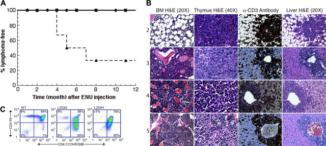
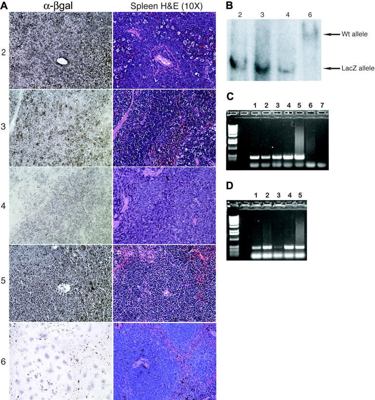
Chromosomal rearrangements affecting RUNX1 and CBFB are common in acute leukemias. These mutations result in the expression of fusion proteins that act dominant-negatively to suppress the normal function of the Runt-related transcription factor 1 (RUNX)/core binding factor beta (CBFbeta) complexes. In addition, loss-of-function mutations in Runt-related transcription factor 1 (RUNX1) have been identified in sporadic cases of acute myeloid leukemia (AML) and in association with the familial platelet disorder with propensity to develop AML (FPD/AML). In order to examine the hypothesis that decreased gene dosage of RUNX1 may be a critical event in the development of leukemia, we treated chimeric mice generated from Runx1(lacZ/lacZ) embryonic stem (ES) cells that have homozygous disruption of the Runx1 gene with N-ethyl-N-nitrosourea (ENU). We observed an increased incidence of T-lymphoblastic lymphoma in Runx1(lacZ/lacZ) compared with wild-type chimeras and confirmed that the tumors were of ES-cell origin. Our results therefore suggest that deficiency of Runx1 can indeed predispose mice to hematopoietic malignancies.


Figures

Figure 1.
Figure 1.
Runx1lacZ/lacZ chimeras are more susceptible to developing T-lymphoblastic lymphoma than wild-type chimeras. (A) Kaplan-Meier survival curves (lymphoma-free) are shown for untreated Runx1lacZ/lacZ (n = 6) and Runx1lacZ/lacZ (n = 6) and wild-type (n = 6) chimeras treated with 100 mg/kg ENU. • indicates untreated Runx1lacZ/lacZ chimeras; ▪, wild-type ES-cell chimeras treated with ENU; ▴, Runx1lacZ/lacZ chimeras treated with ENU. (B) Hematoxylin and eosin (H&E) staining of thymus, bone marrow (BM), and liver from LZD nos. 2 through 5. Immunohistochemistry using an anti-CD3 antibody demonstrates that the malignant cells in the livers of these animals stained positively for CD3. (C) Flow cytometry of thymocytes from LZD no. 2 and LZD no. 4 using antibodies against CD4 and CD8 demonstrates that the majority of cells have an abnormal CD4+/CD8+ phenotype. WT indicates wild type. Numbers in each quadrant indicate the percentage of total thymocytes in that quadrant.
Figure 2.
Figure 2.
T-lymphoblastic lymphoma is of ES-cell origin in Runx1lacZ/lacZ chimeras. (A) Immunohistochemistry using an anti–β-galactosidase antibody demonstrates that the malignant cells in the spleens of LZD nos. 2 through 5 express β-galactosidase and are therefore derived from Runx1lacZ/lacZ ES cells. The normal splenocytes from LZD no. 6 (which did not develop lymphoma) serve as negative controls. (B) Southern blot analysis detects presence of only the Runx1-LacZ knock-in allele in lymphoma-effaced bone marrow from LZD nos. 2 through 4 but only wild-type Runx1 allele from normal bone marrow of LZD no. 6. (C) Genomic PCR using LacZ gene primers and DNA from sorted thymic cells of a Runx1lacZ/lacZ chimera. Lane 1, CD8+; lane 2, CD8+/CD4+; lane 3, CD4-/CD8-; lane 4, CD4+; lane 5, total thymus; lane 6, total thymus from a wild-type mouse; lane 7, no DNA. (D) Genomic PCR using LacZ gene primers and DNA from sorted bone marrow cells of a Runx1lacZ/lacZ chimera. Lane 1, c-kit+/lin-; lane 2, c-kit+/lin+; lane 3, c-kit-/lin+; lane 4, c-kit-/lin-; lane 5, total bone marrow.

References

    1. Castilla LH, Wijmenga C, Wang Q, et al. Failure of embryonic hematopoiesis and lethal hemorrhages in mouse embryos heterozygous for a knocked-in leukemia gene CBFB-MYH11. Cell. 1996;87: 687-696. - PubMed
    1. Yergeau DA, Hetherington CJ, Wang Q, et al. Embryonic lethality and impairment of haematopoiesis in mice heterozygous for an AML1-ETO fusion gene. Nat Genet. 1997;15: 303-306. - PubMed
    1. Okuda T, van Deursen J, Hiebert SW, Grosveld G, Downing JR. AML1, the target of multiple chromosomal translocations in human leukemia, is essential for normal fetal liver hematopoiesis. Cell. 1996;84: 321-330. - PubMed
    1. Wang Q, Stacy T, Binder M, Marin-Padilla M, Sharpe AH, Speck NA. Disruption of the Cbfa2 gene causes necrosis and hemorrhaging in the central nervous system and blocks definitive hematopoiesis. Proc Natl Acad Sci U S A. 1996;93: 3444-3449. - PMC - PubMed
    1. Wang Q, Stacy T, Miller JD, et al. The CBFbeta subunit is essential for CBFalpha2 (AML1) function in vivo. Cell. 1996;87: 697-708. - PubMed

MeSH terms

Substances