

doi: 10.1126/science.1114580.
Epub 2005 Jul 28.
Plague bacteria target immune cells during infection
Affiliations
- PMID: 16051750
- PMCID: PMC3210820
- DOI: 10.1126/science.1114580
Item in Clipboard
Plague bacteria target immune cells during infection
Science.
.
Abstract
The plague is caused by the bacterium Yersinia pestis. Plague bacteria are thought to inject effector Yop proteins into host cells via the type III pathway. The identity of the host cells targeted for injection during plague infection is unknown. We found, using Yop beta-lactamase hybrids and fluorescent staining of live cells from plague-infected animals, that Y. pestis selected immune cells for injection. In vivo, dendritic cells, macrophages, and neutrophils were injected most frequently, whereas B and T lymphocytes were rarely selected. Thus, it appears that Y. pestis disables these cell populations to annihilate host immune responses during plague.
Figures

(A) Y. pestis secretes YopM-Bla, but not GST-Bla, into the medium in a type III machinery (yscU)-dependent manner during in vitro secretion assays. Cultures were separated into supernatant (S) and pellet (P) fractions, and proteins were visualized by SDS-polyacrylamide gel electrophoresis (PAGE) and immunoblotting with antisera to β-lactamase (Bla) and RNA polymerase A (RpoA). (B) HeLa cells were infected with Y. pestis carrying either empty vector (center) or YopM-Bla (right), followed by staining with CCF2-AM and visualization of live cells by fluorescence microscopy. Blue and green fluorescence and differential interference contrast images were overlaid to obtain composite images.
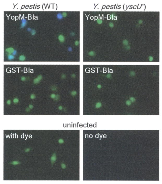
Type III injection of peritoneal macrophages. Peritoneal macrophages were harvested from C57BL/6 mice and infected with Y. pestis strains (wild type or yscU−) carrying either YopM-Bla or GST-Bla. Live cells were stained with CCF2-AM and visualized by fluorescence microscopy.
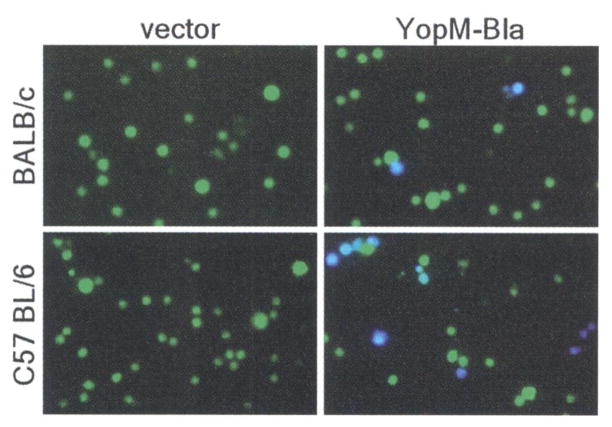
Mice were infected with 105 CFU (BALB/c) or 103 CFU (C57BL/6) of Y. pestis carrying either pHSG576 (vector) or pMM83 (YopM-Bla). After 2 days, infected spleens were removed, homogenized, and incubated with CCF2-AM; live cells were observed by fluorescence microscopy.
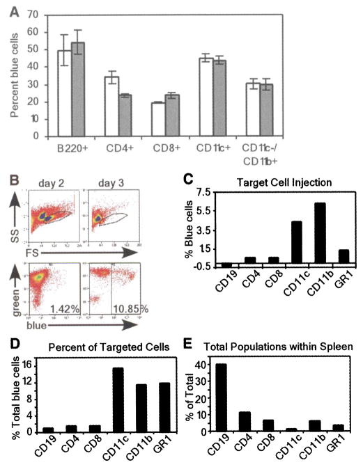
(A) Flow cytometric analysis of in vitro infections of purified cell populations with Y. pestis carrying pMM83 (YopM-Bla, white bars) or pMM85 (YopE-Bla, gray bars). The percentage of live blue cells for each cell type was plotted (data are means ± SEM of triplicate samples). (B) Mice were infected with 103 CFU of Y pestis carrying either pHSG576 (vector) or pMM83 (YopM-Bla). Splenocytes were harvested and stained with CCF2-AM as well as cell-specific surface markers. The R1 gate was set by forward scatter (FS) versus side scatter (SS) and exclusion of propidium iodide–positive cells (top panel). R1-gated cells were analyzed for blue and green fluorescence (bottom panel). (C) APC-conjugated antibodies to cell-specific markers were used to determine percentages of blue cells for each population. (D) The percentage of each cell type in the total population of blue cells was determined by gating on each cell-specific marker. (E) Total population of each cell type in infected spleens.
References
Publication types
MeSH terms
Substances
Grants and funding
LinkOut - more resources
Full Text Sources
Other Literature Sources
Medical
