. 2005 Aug;79(16):10138-46.
doi: 10.1128/JVI.79.16.10138-10146.2005.

RNAs in the virion of Kaposi's sarcoma-associated herpesvirus

Affiliations

RNAs in the virion of Kaposi's sarcoma-associated herpesvirus

Jill Bechtel et al. J Virol. 2005 Aug.

Abstract

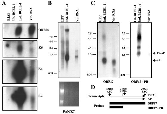
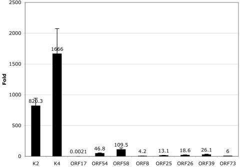
De novo infection of cultured cells with Kaposi's sarcoma-associated herpesvirus (KSHV) typically results in a latent infection. Recently, however, it has been reported that a subset of lytic mRNAs can be detected in cells shortly after KSHV infection; this expression is transient and eventually subsides, leading to latent infection (H. H. Krishnan et al., J. Virol 78:3601-3620, 2004). Since it has been shown that viral RNAs can be packaged into other herpesvirus virions, we sought to determine if KSHV virions contained RNAs and, if so, whether these RNAs contributed to the pool of lytic transcripts detected immediately after infection. Using DNA microarray, reverse transcription (RT)-PCR, and Northern blotting analyses, we identified 11 virally encoded RNAs in KSHV virions. These corresponded in size to the full-length mRNAs found in cytoplasmic RNA, and at least one was directly demonstrated to be translated upon infection in the presence of actinomycin D. Ten of these RNAs correspond to transcripts reported by Krishnan et al. at early times of infection, representing ca. 30% of such RNAs. Thus, import of RNAs in virions can account for some but not all of the early-appearing lytic transcripts. Quantitative RT-PCR analysis of infected-cell RNA demonstrated that most of the virion RNAs were very abundant at late times of infection, consistent with nonspecific incorporation during budding. However, the intracellular levels of one virion mRNA, encoding the viral protease, were much lower than those of transcripts not packaged in the virus particle, strongly suggesting that it may be incorporated by a specific mechanism.


Figures

FIG. 1.
FIG. 1.
Identification of KSHV virion RNAs by DNA array. RNAs isolated from BJAB cells and KSHV virions were reverse transcribed in the presence of amino-acyl dNTPs and then coupled to Cy3 and Cy5 dyes, respectively. The labeled cDNAs were then hybridized to the KSHV tile array as described in the text. The histogram displays the virion RNA signals across the KSHV tile array. The line across the histogram represents the arbitrary cutoff used in our analysis. The labeled peaks represent the RNAs consistently found in our KSHV virions.
FIG. 2.
FIG. 2.
RT-PCR confirms the presence of KSHV virion RNAs. RNAs isolated from BJAB cells, uninduced and induced BCBL-1 cells, and KSHV virions were reverse transcribed using an oligo(dT) primer and then subjected to PCR with gene-specific primers. The PCR products were separated on 1.5 to 2% agarose gels. Lanes 1, KSHV BAC DNA; lanes 2, uninduced BCBL-1 cDNA; lanes 3, induced BCBL-1 cDNA; lanes 4, KSHV virion cDNA. Molecular size standards are the GeneRuler 100-bp DNA Ladder (Fermentas).
FIG. 3.
FIG. 3.
Northern blots of KSHV virion RNA confirm that the transcripts are full length. (A) KSHV virion (Vir.) RNA and poly(A)-purified RNAs isolated from BJAB cells and uninduced (Un.) and induced (Ind.) BCBL-1 cells were separated on a 1% formaldehyde agarose gel and then blotted to nylon membranes and probed for ORF54, K6, K4, and K2 using strand-specific riboprobes. (B) RNAs isolated from HFFs, induced BCBL-1 cells, and KSHV virions were separated on a 1% formaldehyde agarose gel, transferred to nylon, and then probed for PAN (double-stranded DNA probe). The ethidium bromide staining of the Northern blot is shown below. (C) RNAs isolated from HFFs, induced BCBL-1 cells, and KSHV virions were separated on a 1% formaldehyde agarose gel, transferred to nylon, and then probed with a riboprobe to the entire ORF17 locus. Virion RNA and poly(A)-purified RNAs from uninduced and induced BCBL-1 cells were separated on a 1% agarose gel, transferred to nylon, and then hybridized with a riboprobe specific to the protease portion of ORF17. PR/AP and AP transcripts are marked with arrows. (D) Schematic diagram of two transcripts produced from the ORF17 locus. The start and stop codons for the longer PR/AP transcript are indicated, as are the two known transcriptional start sites for the AP transcript. The numbers refer to the genomic map positions. The two boxes represent the regions used as riboprobes in the Northern blots shown in Fig. 3C. The RNA ladders depicted in panels B and C are in kb.
FIG. 4.
FIG. 4.
ORF59 mRNA packaged into virions is competent for translation. 293 cells were mock or KSHV infected in the presence of DMSO, actinomycin D (2 μg/ml), or cycloheximide (100 μg/ml) and labeled continuously with [35S]methionine and cysteine under these conditions for either 4 or 8 h. The cells were washed and lysed in 1% Ipegal buffer and then subjected to immunoprecipitation with antibodies specific for ORF59 (A) or LANA (B). (A) 35S-labeled ORF59 immunoprecitates were separated on a 10% SDS-PAGE gel, fixed, treated with Enhance (Perkin-Elmer), and then dried and exposed to film. The arrow indicates the ORF59 protein. (B) 35S-labeled LANA immunoprecipitates were separated on a 7.5% SDS-PAGE gel and then transferred to polyvinylidene difluoride and probed for LANA. MD, mock infected with DMSO; MA, mock infected with actinomycin D; MC, mock infected with cycloheximide; 4KA and 8KA, KSHV infected for 4 or 8 h with actinomycin D; KD, KSHV infected with DMSO; KC, KSHV infected with cycloheximide. Cycloheximide samples were harvested at 4 h postinduction. DMSO and actinomycin D samples were harvested at 8 h postinduction unless otherwise noted.
FIG. 5.
FIG. 5.
Real-time PCR suggests that the ORF17 RNA is specifically incorporated into KSHV virions. RNA from induced BCBL-1 cells (3 days postinoculation) was reverse transcribed with an oligo(dT) primer and subjected to real-time PCR using gene-specific primers. Standard curves were generated for each primer/probe set (listed in Table 2) and then used to calculate the relative amounts of transcripts present in the sample. Levels of KSHV transcripts were normalized to levels of GAPDH transcript. The graph displays the normalized levels of each transcript (n-fold) over GAPDH. The data values are listed with the corresponding bars, and the error bars representing the standard deviations are shown.

References

    1. Akula, S. M., P. P. Naranatt, N. S. Walia, F. Z. Wang, B. Fegley, and B. Chandran. 2003. Kaposi's sarcoma-associated herpesvirus (human herpesvirus 8) infection of human fibroblast cells occurs through endocytosis. J. Virol. 77:7978-7990. - PMC - PubMed
    1. Akula, S. M., N. P. Pramod, F. Z. Wang, and B. Chandran. 2002. Integrin α3β1 (CD 49c/29) is a cellular receptor for Kaposi's sarcoma-associated herpesvirus (KSHV/HHV-8) entry into the target cells. Cell 108:407-419. - PubMed
    1. Ballestas, M. E., P. A. Chatis, and K. M. Kaye. 1999. Efficient persistence of extrachromosomal KSHV DNA mediated by latency-associated nuclear antigen. Science 284:641-644. - PubMed
    1. Bechtel, J. T., Y. Liang, J. Hvidding, and D. Ganem. 2003. Host range of Kaposi's sarcoma-associated herpesvirus in cultured cells. J. Virol. 77:6474-6481. - PMC - PubMed
    1. Bechtel, J. T., R. C. Winant, and D. Ganem. 2005. Host and viral proteins in the virion of Kaposi's sarcoma-associated herpesvirus. J. Virol. 79:4952-4964. - PMC - PubMed

MeSH terms

LinkOut - more resources