. 2005 Aug;79(16):10155-63.
doi: 10.1128/JVI.79.16.10155-10163.2005.

Envelope targeting: hemagglutinin attachment specificity rather than fusion protein cleavage-activation restricts Tupaia paramyxovirus tropism

Affiliations

Envelope targeting: hemagglutinin attachment specificity rather than fusion protein cleavage-activation restricts Tupaia paramyxovirus tropism

Christoph Springfeld et al. J Virol. 2005 Aug.

Abstract

To engineer a targeting envelope for gene and oncolytic vector delivery, we characterized and modified the envelope proteins of Tupaia paramyxovirus (TPMV), a relative of the morbilli- and henipaviruses that neither infects humans nor has cross-reactive relatives that infect humans. We completed the TPMV genomic sequence and noted that the predicted fusion (F) protein cleavage-activation site is not preceded by a canonical furin cleavage sequence. Coexpression of the TPMV F and hemagglutinin (H) proteins induced fusion of Tupaia baby fibroblasts but not of human cells, a finding consistent with the restricted TPMV host range. To identify the factors restricting fusion of non-Tupaia cells, we initially analyzed F protein cleavage. Even without an oligo- or monobasic protease cleavage sequence, TPMV F was cleaved in F1 and F2 subunits in human cells. Edman degradation of the F1 subunit yielded the sequence IFWGAIIA, placing the conserved phenylalanine in position 2, a novelty for paramyxoviruses but not the cause of fusion restriction. We then verified whether the lack of a TPMV H receptor limits fusion. Toward this end, we displayed a single-chain antibody (scFv) specific for the designated receptor human carcinoembryonic antigen on the TPMV H ectodomain. The H-scFv hybrid protein coexpressed with TPMV F mediated fusion of cells expressing the designated receptor, proving that the lack of a receptor limits fusion and that TPMV H can be retargeted. Targeting competence and the absence of antibodies in humans define the TPMV envelope as a module to be adapted for ferrying ribonucleocapsids of oncolytic viruses and gene delivery vectors.


Figures
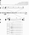
FIG. 1.
FIG. 1.
TPMV genome organization and glycoproteins structure and sequence. (A) Comparison of the H protein cytoplasmic tails of selected paramyxoviruses. The proteins are aligned based on a charged amino acid postulated to be the first cytoplasmic residues. Amino acids in the transmembrane region are italicized. The number of the last amino acid in each sequence is indicated on the right. (B) Schematic drawing of the TPMV H protein. The cytoplasmic tail (CT), transmembrane region (TM), and three predicted N glycosylation sites (▾) are indicated. (C) Map of the TPMV genome. The black bar represents the 17,904-nucleotide single-stranded TPMV antigenome; the eight open arrows indicate the position of the ORFs of the N, P, C, V, M, F, H, and L proteins; and the small vertical bars indicate the positions of the intergenic trinucleotides. (D) Schematic drawing of the TPMV F protein. SP, signal peptide; FP, fusion peptide; TM, transmembrane region; CT, cytoplasmic tail. The black arrow indicates the cleavage site situated between aa 107 and 108; the black triangles (▾) indicate the N glycosylation sites. (E) Alignment of the F2-F1 junction of selected paramyxoviruses. Identical amino acids are indicated by an asterisk, highly conserved amino acids are indicated by a colon, and well conserved amino acids are indicated by a period. Conserved basic amino acids and the phenylalanine/leucine conserved as position one of the fusion peptide in the majority of paramyxoviruses are in boldface. The number of the last amino acid in each sequence is indicated on the right. Abbreviations not introduced in the text: MoV, Mossman virus; HeV, Hendra virus; RPV, Rinderpest virus; CDV, canine distemper virus; PDV, phocine distemper virus; hPIV1, human parainfluenza virus type 1; hPIV3, human parainfluenza virus type 3; SV5, simian virus 5. The number of the last amino acid in each sequence is indicated on the right.
FIG. 2.
FIG. 2.
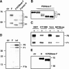
Western blot analysis of the TPMV glycoproteins. (A) TPMV F protein expressed in TBF cells, fractionated by SDS-10% PAGE, and detected with a peptide antiserum against the cytoplasmic tail. The two bands correspond to the TPMV F0 and F1 fragments. (B) Deglycosylation of the TPMV F0 fragment with increasing concentrations of PNGase F (from left to right) results in five distinct bands, confirming that all four potential N glycosylation sites are used. (C) Cleavage of the TPMV F protein in cells from three species. TPMV F was transiently expressed in different cells lines, fractionated by SDS-10% PAGE, and detected with the anti-F peptide antiserum. The positions of the F0 and F1 fragments are indicated. (D) TPMV F is cleaved membrane proximally, resulting in an F1b fragment. Protein lysates from transfected (central lane) and infected (right lane) TBF cells were fractionated by SDS-15% PAGE, and F proteins were detected using by a peptide antiserum against the cytoplasmic tail. The positions of the F0, F1, and F1b fragments are indicated on the right; the positions of molecular mass markers are indicated on the left in kilodaltons. (E) Expression of the TPMV H protein in 293T cells and glycosylation analysis. Protein extracts of control cells (first lane) or transfected cells treated or not with PNGase F (second and third lanes) were loaded, and the H protein was detected with a peptide antiserum against the cytoplasmic tail.
FIG. 3.
FIG. 3.
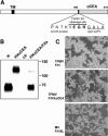
The TPMV glycoproteins induce syncytium formation in TBF cells. TBF cells were transfected with a plasmid encoding the F protein alone (upper panel) or with plasmids encoding both TPMV glycoproteins (lower panel) and photographed after 16 h. Coexpression of TPMV F and H results in the formation of multinucleated syncytia.
FIG. 4.
FIG. 4.
Retargeting of the TPMV H protein. (A) Schematic drawing of the hybrid protein composed of TPMV H and the scFv against CEA. The positions of the transmembrane domain (TM) and the factor Xa cleavage site are indicated by black boxes. In the lower part of the panel the amino acid sequence of the junction is shown. (B) TPMV H and HXαCEA proteins transiently expressed in 293 cells. The anti-CEA scFv can be cleaved off with factor Xa (right lane). (C) Expression of TPMV F and TPMV HXαCEA in MC38cea cells results in the formation of syncytia (central panel). The standard MV F protein was coexpressed with a retargeted MV H protein (MV-F/HXL) (15) as a positive control (lower panel). The standard TPMV H protein was unable to fuse murine cells when cotransfected with TPMV F (upper panel).

References

    1. Asada, T. 1974. Treatment of human cancer with mumps virus. Cancer 34:1907-1928. - PubMed
    1. Bahr, U., and G. Darai. 2001. Analysis and characterization of the complete genome of tupaia (tree shrew) herpesvirus. J. Virol. 75:4854-4870. - PMC - PubMed
    1. Bell, J. C., B. Lichty, and D. Stojdl. 2003. Getting oncolytic virus therapies off the ground. Cancer Cell 4:7-11. - PubMed
    1. Bendtsen, J. D., H. Nielsen, G. von Heijne, and S. Brunak. 2004. Improved prediction of signal peptides: SignalP 3.0. J. Mol. Biol. 340:783-795. - PubMed
    1. Bucheit, A. D., S. Kumar, D. M. Grote, Y. Lin, V. von Messling, R. B. Cattaneo, and A. K. Fielding. 2003. An oncolytic measles virus engineered to enter cells through the CD20 antigen. Mol. Ther. 7:62-72. - PubMed

Publication types

LinkOut - more resources