

High-resolution computed tomography imaging of airway disease in infants with cystic fibrosis
- PMID: 16051903
- PMCID: PMC2718397
- DOI: 10.1164/rccm.200412-1665OC
High-resolution computed tomography imaging of airway disease in infants with cystic fibrosis
Abstract
Rationale: The development of early lung disease in patients with cystic fibrosis (CF) remains poorly defined.
Objective: Determine whether asymptomatic infants with CF have evidence for changes in airway structure when assessed by high-resolution computed tomography, and whether airway structure correlates with airway function in this age group.
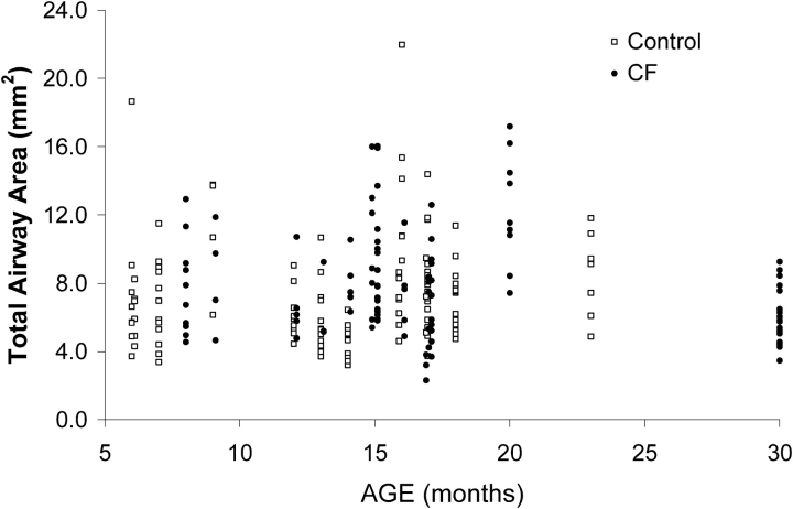
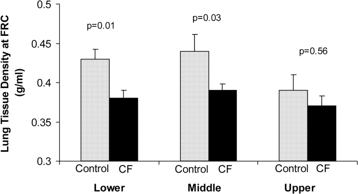
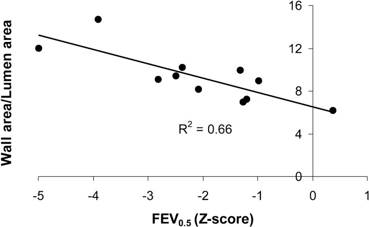
Methods: Thirteen infants with CF (8-33 mo) and 13 control infants (7-25 mo) were evaluated. Airway wall and lumen areas were measured from three 1-mm-thick cross-sectional images obtained from upper, middle, and lower lobes during a respiratory pause with the lungs inflated to an airway pressure of 20 cm H2O. Lung tissue density was measured from images obtained during a respiratory pause at FRC. Forced expiratory flows were measured by the rapid thoracic compression technique in 11 infants with CF.
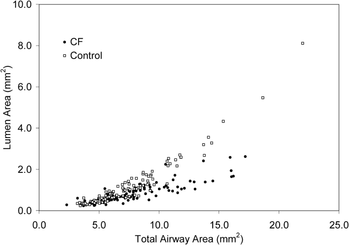
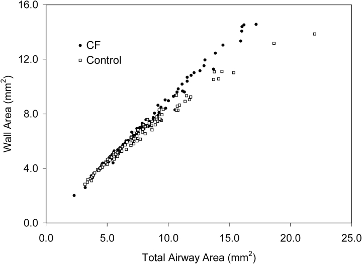
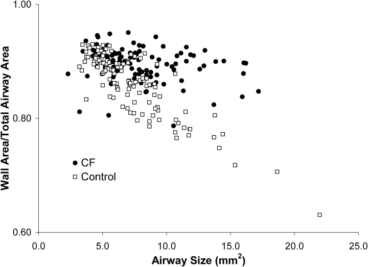
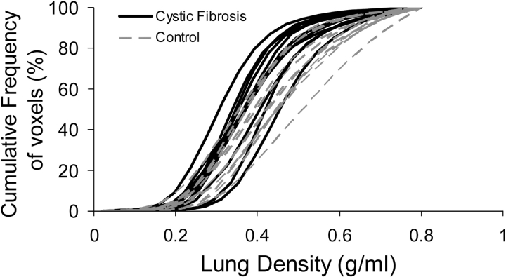
Results: Airway wall area increased more per unit increase in airway size, whereas airway lumen area increased less per unit increase in airway size in the CF than in the control group. Among infants with CF, a greater ratio of wall to lumen area correlated with lower airway function. In addition, lung density at relaxed (passive) FRC was lower for infants with CF than for control infants (0.38 vs. 0.43 g/ml; p < 0.02).
Conclusions: Our results indicate that infants with CF have thickened airway walls, narrowed airway lumens, and air trapping, when assessed by high-resolution computed tomography, and measurements of airway structure correlated with airway function.
Figures
References
-
- Tepper RS, Montgomery GL, Ackerman V, Eigen H. Longitudinal evaluation of pulmonary function in infants and very young children with cystic fibrosis. Pediatr Pulmonol 1993;16:96–100. - PubMed
-
- Ranganathan SC, Dezateux C, Bush A, Carr SB, Castle RA, Madge S, Price J, Stroobant J, Wade A, Wallis C, et al., and London Collaborative Cystic Fibrosis Group. Airway function in infants newly diagnosed with cystic fibrosis. Lancet 2001;358:1964–1965. - PubMed
-
- Tepper RS, Hiatt P, Eigen H, Scott P, Grosfeld J, Cohen M. Infants with cystic fibrosis: pulmonary function at diagnosis. Pediatr Pulmonol 1988;5:15–18. - PubMed
-
- Ranganathan SC, Bush A, Dezateux C, Carr SB, Hoo AF, Lum S, Madge S, Price J, Stroobant J, Wade A, et al. Relative ability of full and partial forced expiratory maneuvers to identify diminished airway function in infants with cystic fibrosis. Am J Respir Crit Care Med 2002;166:1350–1357. - PubMed
-
- Khan TZ, Wagener JS, Bost T, Martinez J, Accurso FJ, Riches DW. Early pulmonary inflammation in infants with cystic fibrosis. Am J Respir Crit Care Med 1995;151:1075–1082. - PubMed
