. 2006 Jan 11;25(1):1-12.
doi: 10.1038/sj.emboj.7600759. Epub 2005 Jul 28.

EGF stimulates annexin 1-dependent inward vesiculation in a multivesicular endosome subpopulation

Affiliations

EGF stimulates annexin 1-dependent inward vesiculation in a multivesicular endosome subpopulation

Ian J White et al. EMBO J. .

Abstract

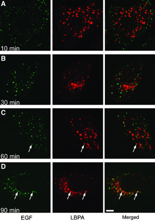
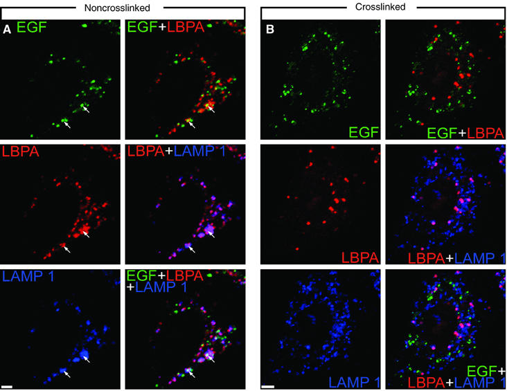
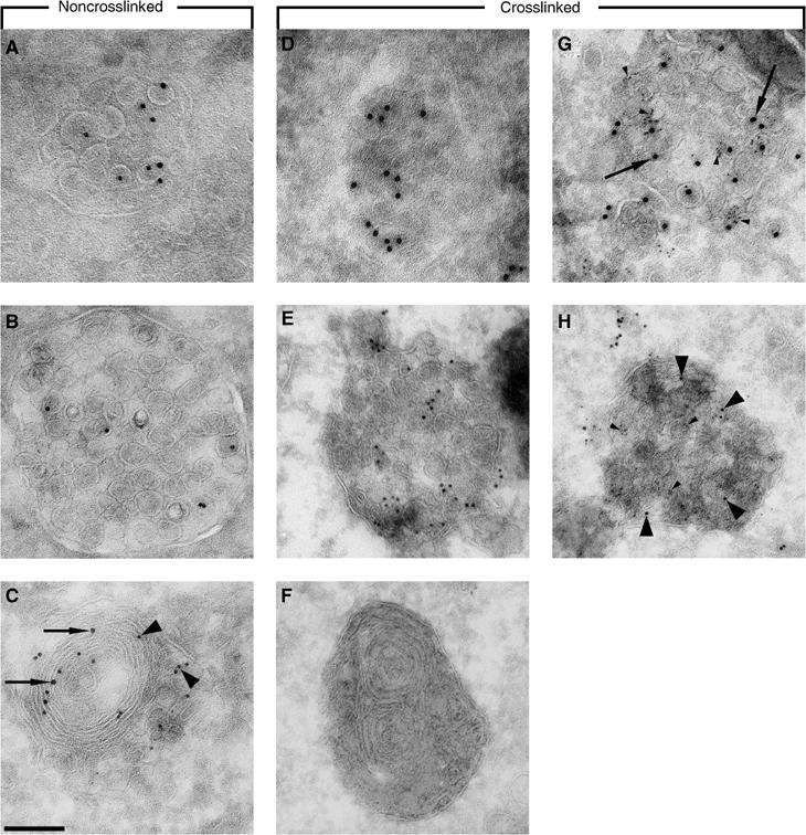
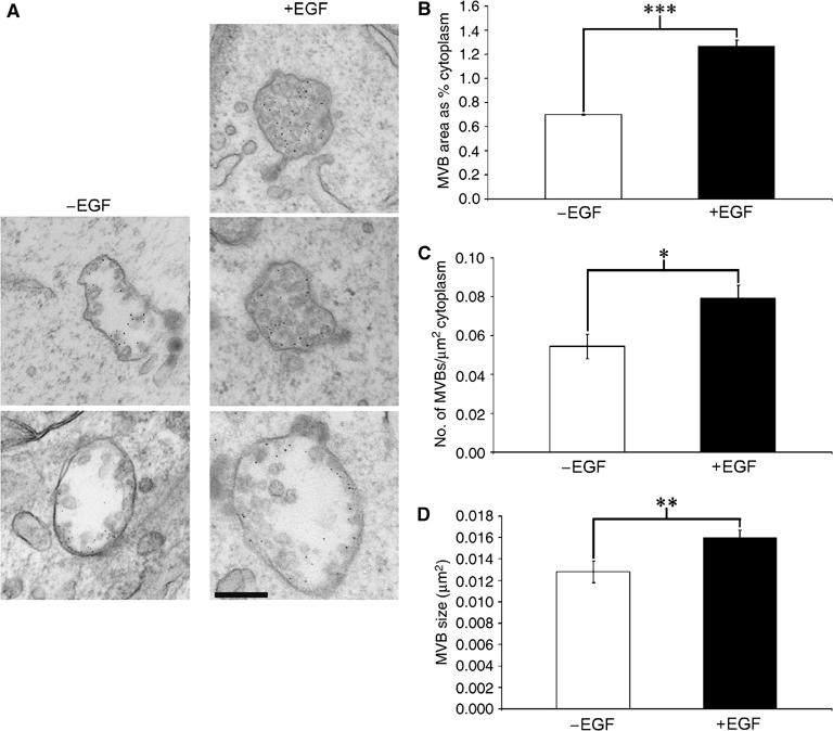
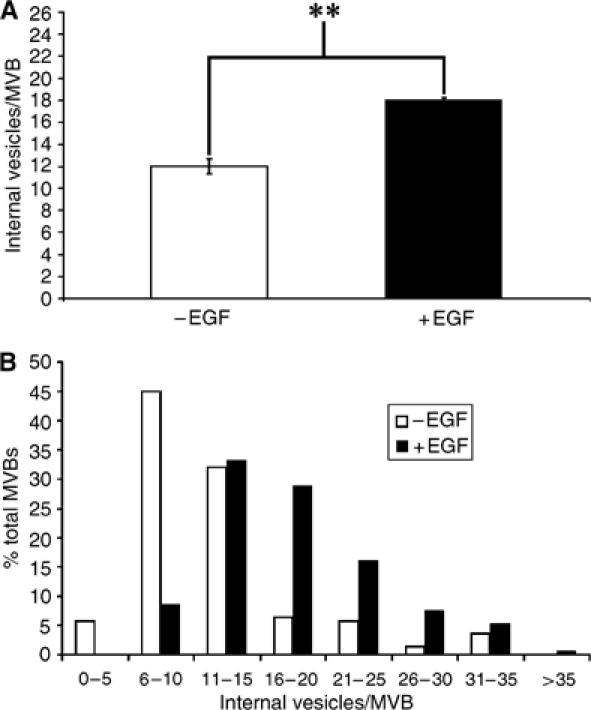
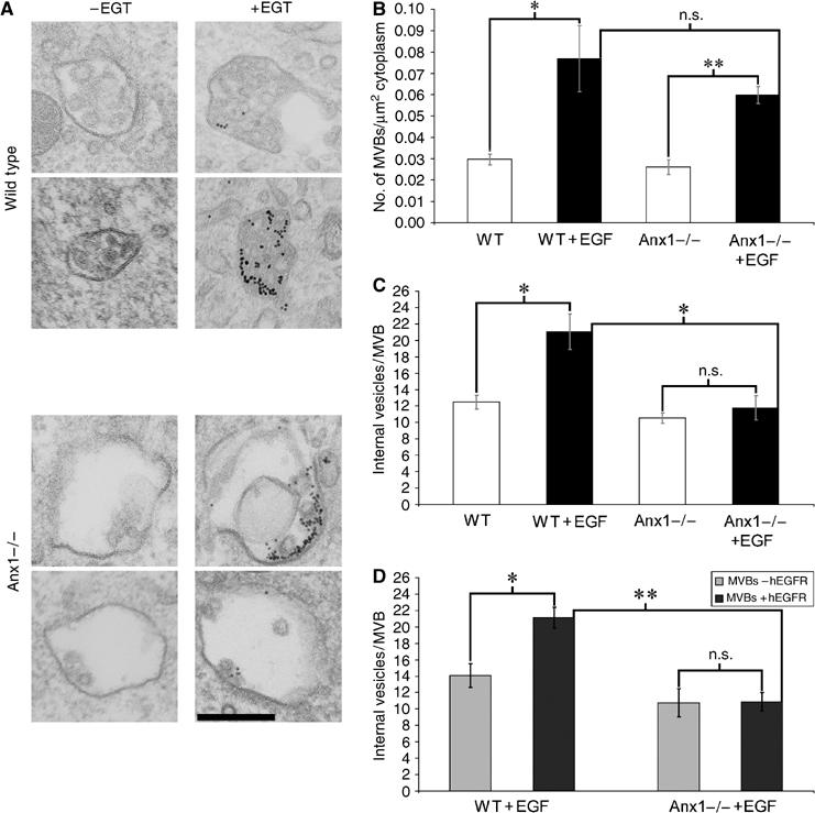
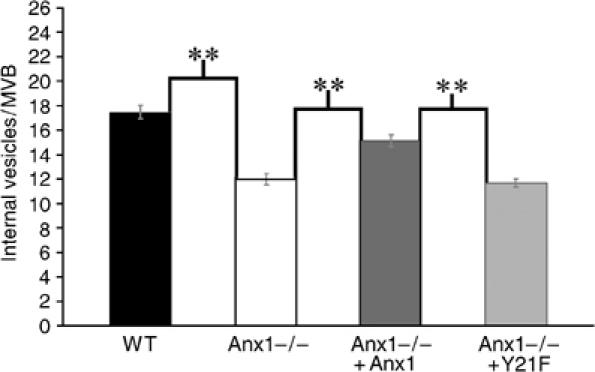
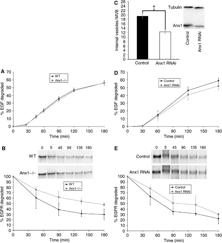
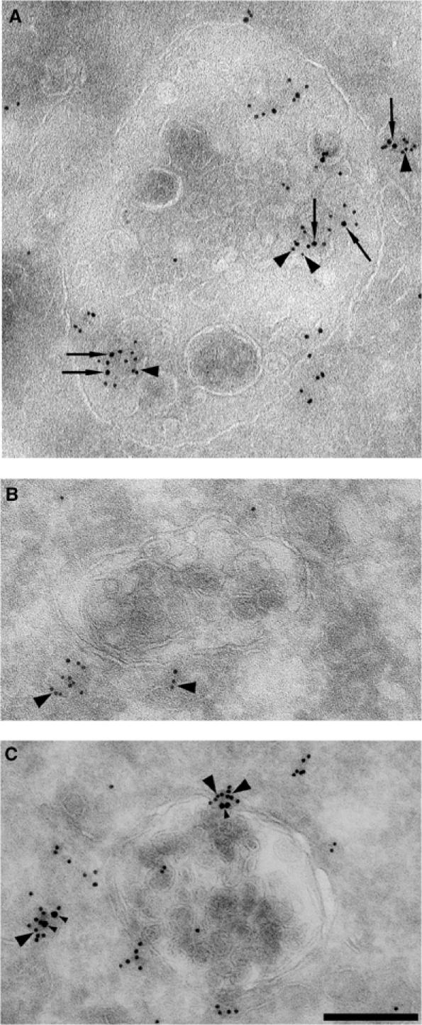
Here we show that EGF and EGF receptor (EGFR) are trafficked through a subpopulation of multivesicular endosomes/bodies (MVBs) that are distinct from morphologically identical vacuoles that label for the late endosomal marker lyso-bisphosphatidic acid (LBPA). EGF stimulation increases both MVB biogenesis and inward vesiculation within EGFR-containing MVBs. Deletion of annexin 1, a substrate of EGFR tyrosine kinase, abolishes the effect of EGF stimulation on inward vesiculation. This phenotype is reversible by transfection with wild-type but not Y21F phosphorylation mutant annexin 1. Deletion of annexin 1 has no effect on EGF-stimulated MVB biogenesis, suggesting that MVB biogenesis and inward vesiculation within MVB are mediated by separate mechanisms. Loss or depletion of annexin 1 has no effect on EGF degradation and causes only a small delay in EGFR degradation, indicating that annexin 1 operates downstream of Hrs- and ESCRT-mediated sorting and is required solely for EGF-stimulated inward vesiculation. Annexin 1 accumulates on internal vesicles of MVB after EGF-stimulated inward vesiculation, suggesting that it may be required for a late stage in inward vesiculation.


Figures

Figure 1
Figure 1
EGF does not enter LBPA-positive compartments till late in the endocytic pathway. HEp2 cells were stimulated with fluorescent EGF for the indicated times A=10 min, B=30 min, C=60 min, and D=90 min. Cells were fixed prior to permeabilisation and then stained with an antibody against LBPA. Colocalisation between EGF and LBPA (arrows) appears as yellow in the merged image. Colocalisation was limited and observed only after 60 and 90 min. Bar=5 μm.
Figure 2
Figure 2
Inhibition of MVB–lysosome fusion inhibits delivery of EGF to LBPA-positive lysosomes. HEp2 cells were loaded with HRP in preparation for crosslinking. Noncrosslinked cells treated without DAB/H2O2 were stimulated with fluorescent EGF at 37°C for 60 min (A). Cells that were crosslinked with DAB/H2O2 were stimulated with fluorescent EGF for 180 min at 37°C (B). Cells were fixed prior to permeabilisation and then stained with antibodies against LBPA and LAMP 1. Colocalisation between EGF and LBPA appears as yellow in the merged images, LBPA and LAMP 1 as magenta, and colocalisation between all three (examples shown by arrows) as white. Vacuoles staining for both EGF and LBPA in noncrosslinked cells always also stain for LAMP 1. Crosslinking of lysosomes abolishes both the majority of LBPA staining and any colocalisation between EGF and LBPA. Bars=5 μm.
Figure 3
Figure 3
EGFR and LBPA appear in distinct MVBs that also contain CD63, and colocalise only in lysosomes. HEp2 cells were loaded with HRP in preparation for crosslinking. Noncrosslinked cells treated without DAB/H2O2 (A–C) were stimulated with EGF for 60 min at 37°C and those that were crosslinked with DAB/H2O2 (D–H) were stimulated with EGF for 180 min at 37°C. Cells were prepared for cryo-immuno-EM and ultrathin frozen sections either double labelled for EGFR with 15 nm gold (arrows), and LBPA with 10 nm gold (arrowheads) (A–F), or triple labelled for EGFR and LBPA as before, and also for CD63 with 5 nm gold (small arrowheads) (G, H). Panels A and D show MVBs labelling for EGFR, panels B and E MVBs labelling for LBPA and panels C and F lysosomes. Note that in the noncrosslinked cell, the lysosome labels for both EGFR and LBPA, whereas lysosomes in the crosslinked sample label for neither. Panels G and H show that MVBs containing EGFR (G) and those containing LBPA (H) also label for CD63. Bar=200 nm.
Figure 4
Figure 4
EGF stimulates the formation of MVBs. HEp2 cells with lysosomes crosslinked were incubated in the presence or absence of EGF for 60 min at 37°C in the presence of 10 nm BSA gold. (A) Electron micrographs of MVBs from both EGF-stimulated and nonstimulated samples. (B) Mean total MVB area as a percentage of total cytoplasmic area. (C) Mean numbers of MVBs/unit area cytoplasm. (D) Mean MVB sizes. There are significant increases in total area and frequency of MVBs, and a small but significant increase in MVB size upon EGF stimulation. *P<0.05, **P<0.01, ***P<0.001. Data shown are the mean±s.e.m. of three independent experiments. Bar=200 nm.
Figure 5
Figure 5
EGF stimulates the formation of internal vesicles within MVBs. HEp2 cells with lysosomes crosslinked were incubated in the presence or absence of EGF for 60 min at 37°C in the presence of 10 nm BSA gold. Sections were analysed by EM. Internal vesicle numbers were recorded from MVBs chosen randomly over three independent experiments. (A) Mean numbers of internal vesicles/MVB ±s.e.m. (B) Percentage of total MVBs containing different numbers of internal vesicles. **P<0.01. Internal vesicle numbers are significantly increased upon stimulation with EGF.
Figure 6
Figure 6
In annexin 1 knockout cells, EGF stimulates MVB formation, but not internal vesicle formation within MVBs. Wild-type (WT) and annexin 1 knockout (Anxl−/−) cells were transfected with hEGFR. Cells were incubated in the absence or presence of EGF, and hEGFR antibody conjugated to 10 nm gold for 60 min at 37°C. (A) Electron micrographs of MVBs from both EGF-stimulated and nonstimulated samples in wild-type and annexin 1 knockout cells. (B) Mean number of MVBs per unit area of cytoplasm. (C) Mean numbers of internal vesicles/MVB. (D) Mean numbers of internal vesicles/MVB from EGF–stimulated successfully hEGFR-transfected cells only. Mean numbers of internal vesicles are shown in MVBs either containing or not containing hEGFR. *P<0.05, **P<0.01, n.s., no significant difference. Data shown are the mean±s.e.m. of three independent experiments. Annexin 1 knockout does not have a significant effect on the EGF-stimulated increase in MVB formation, but does prevent the EGF-stimulated increase in MVB internal vesicle numbers. The effect of EGF on internal vesicle formation in wild-type cells is limited to MVBs containing hEGFR. Bar=200 nm.
Figure 7
Figure 7
In annexin 1 knockout cells, ectopic expression of annexin 1, but not an annexin 1 tyrosine phosphorylation mutant, partially rescues the EGF-stimulated internal vesicle increase phenotype. Wild-type (WT), annexin 1 knockout cells (Anxl−/−), annexin 1 knockout cells transfected with annexin 1-GFP (Anxl−/− +Anxl) or a Y21F-GFP tyrosine phosphorylation mutant of annexin 1 (Anxl−/− +Y21F) were incubated for 60 min at 37°C in the presence of EGF and 10 nm BSA gold. Sections were analysed by EM. The figure shows mean numbers of internal vesicles/MVB ±s.e.m. of three independent experiments, **P<0.01. Expression of annexin 1-GFP in annexin 1 knockout cells caused a significant increase in MVB internal vesicle count partially rescuing the phenotype seen in wild-type cells. Expression of Y21F-GFP had no significant effect on the number of internal vesicles found when compared to untransfected knockout cells.
Figure 8
Figure 8
Annexin 1 depletion has no effect on EGF degradation, and causes only minor delays in EGFR degradation. Wild-type and annexin 1 knockout mouse lung fibroblast cells (A, B) and HeLa cells treated with control siRNA or annexin 1 siRNA (C–E) were incubated with 125I-labelled EGF (1 ng/ml) for 10 min and chased for the times shown with serum-free media, and the % EGF degradation determined (A, D). The degradation of EGFR was also measured by immunoblotting for the presence of EGFR at the time points illustrated (B, E). All data points are mean±s.e.m. of three independent experiments. HeLa cells treated with annexin 1 siRNA showed reduced annexin 1 levels and display a similar phenotype to annexin 1 knockout cells in that EGF-stimulated inward vesiculation is blocked (C). Annexin 1 knockout or depletion has no effect on the levels or kinetics of EGF degradation, and induces only a small delay in the degradation of EGFR.
Figure 9
Figure 9
EGF stimulation causes annexin 1 to relocalise to MVBs containing EGFR. HEp2 cells stably expressing annexin l-GFP were incubated in the absence of EGF (C) or for 30 min at 37°C in the presence of EGF (A, B). Cells were prepared for cryo-immuno-EM and ultrathin frozen sections either double labelled for EGFR with 15 nm gold (arrows), and GFP with 10 nm gold (large arrowheads) (A and B), or double labelled for transferrin receptor with 15 nm gold (small arrowheads), and GFP with 10 nm gold (large arrowheads) (C). In the absence of EGF stimulation, annexin 1-GFP is found at the plasma membrane, or associated with transferrin receptor in small vesicles, or on the perimeter membrane of MVBs (C). Following EGF stimulation, annexin 1 localises to those MVBs that contain EGFR, where it is found on EGFR-containing internal vesicles (A), but not MVBs that do not contain EGFR (B).

Similar articles

Cited by

References

    1. Babst M, Katzmann DJ, Estepa-Sabal EJ, Meerloo T, Emr SD (2002a) Escrt-III: an endosome-associated heterooligomeric protein complex required for mvb sorting. Dev Cell 3: 271–282 - PubMed
    1. Babst M, Katzmann DJ, Snyder WB, Wendland B, Emr SD (2002b) Endosome-associated complex, ESCRT-II, recruits transport machinery for protein sorting at the multivesicular body. Dev Cell 3: 283–289 - PubMed
    1. Babst M, Odorizzi G, Estepa EJ, Emr SD (2000) Mammalian tumor susceptibility gene 101 (TSG101) and the yeast homologue, Vps23p, both function in late endosomal trafficking. Traffic 1: 248–258 - PubMed
    1. Bache KG, Brech A, Mehlum A, Stenmark H (2003) Hrs regulates multivesicular body formation via ESCRT recruitment to endosomes. J Cell Biol 162: 435–442 - PMC - PubMed
    1. Bache KG, Slagsvold T, Cabezas A, Rosendal KR, Raiborg C, Stenmark H (2004) The growth-regulatory protein HCRPl/hVps37A is a subunit of mammalian ESCRT-I and mediates receptor downregulation. Mol Biol Cell 15: 4337–4346 - PMC - PubMed

Publication types

MeSH terms