. 2005 Aug 8;93(3):319-30.
doi: 10.1038/sj.bjc.6602720.

Fluvastatin synergistically enhances the antiproliferative effect of gemcitabine in human pancreatic cancer MIAPaCa-2 cells

Affiliations

Fluvastatin synergistically enhances the antiproliferative effect of gemcitabine in human pancreatic cancer MIAPaCa-2 cells

G Bocci et al. Br J Cancer. .

Abstract

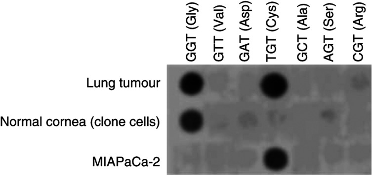
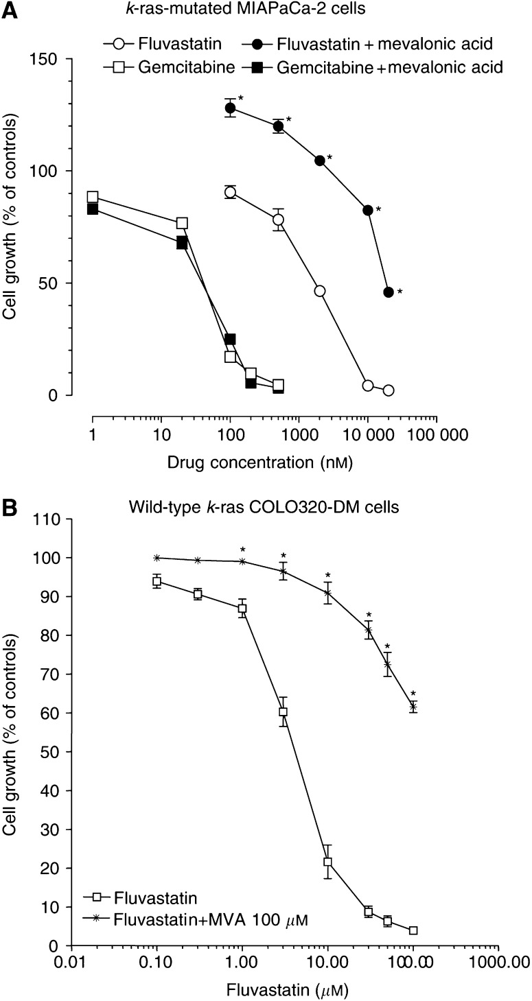
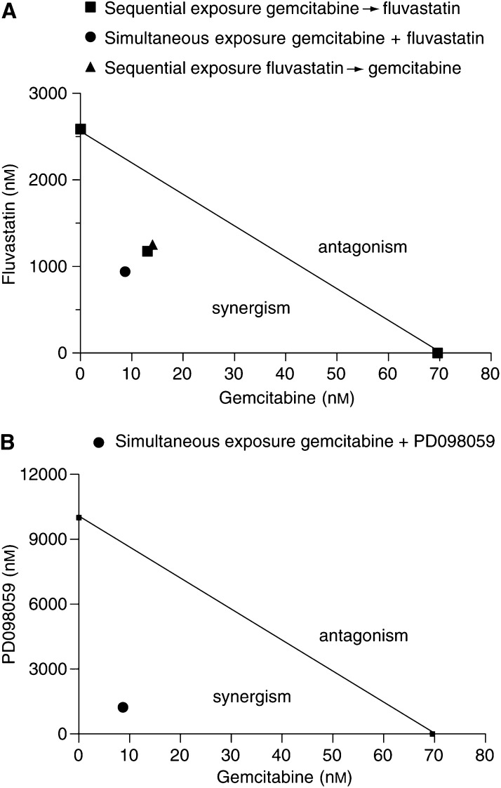
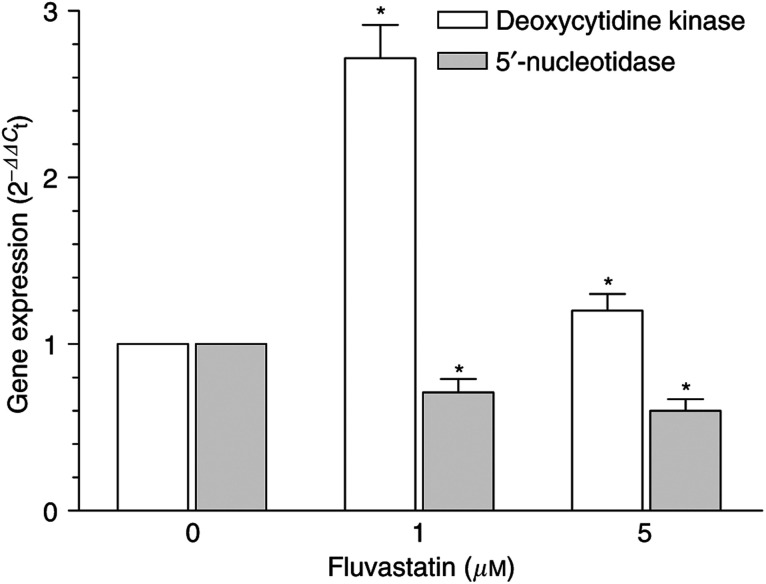
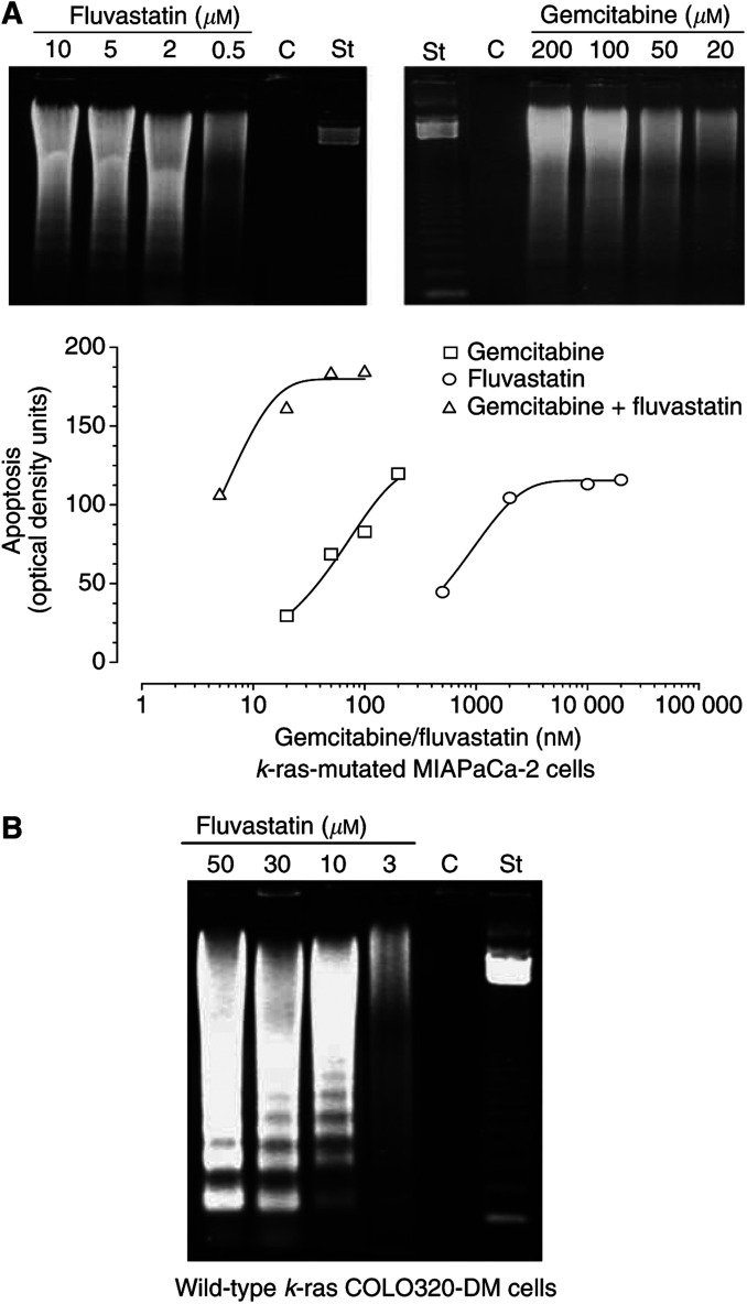
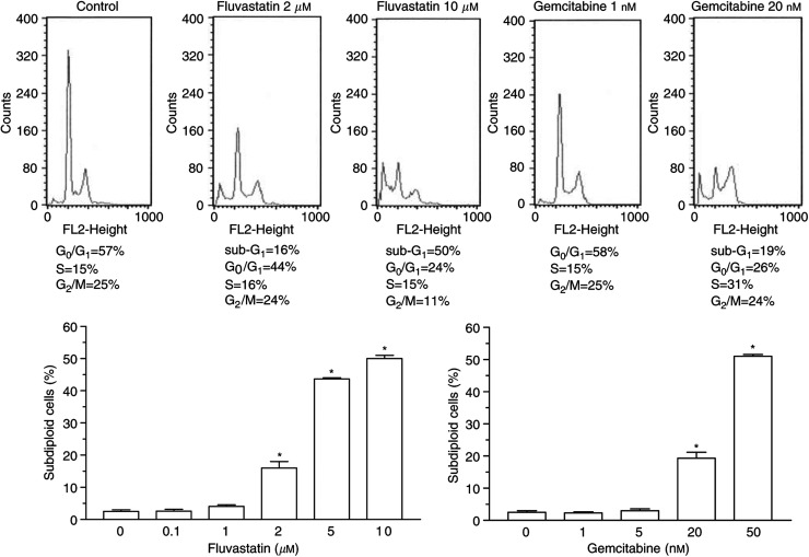
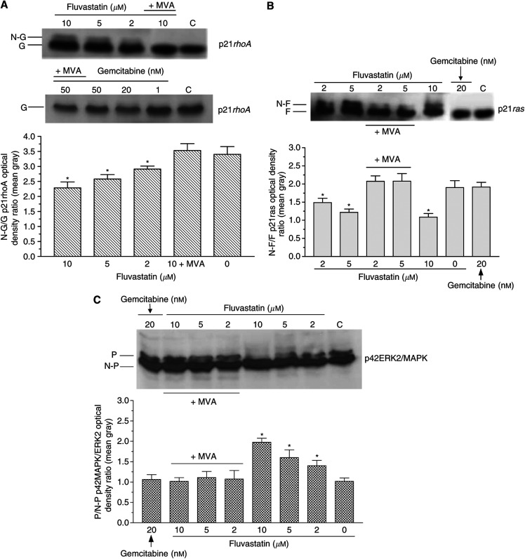
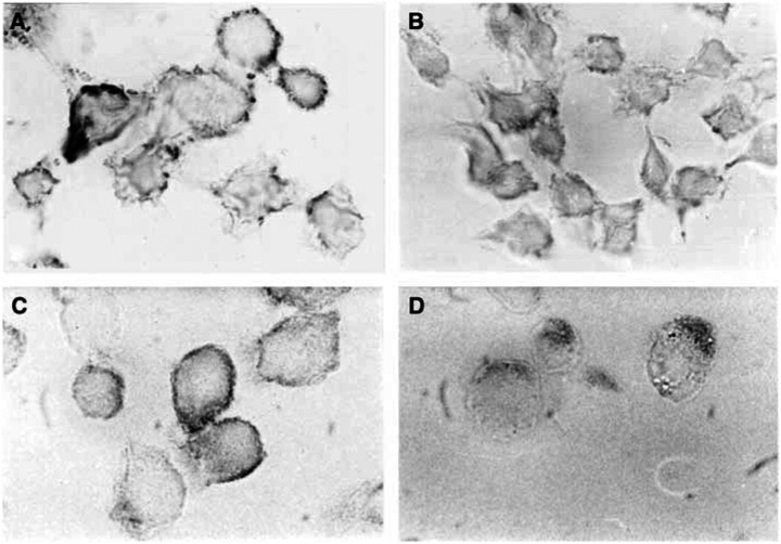
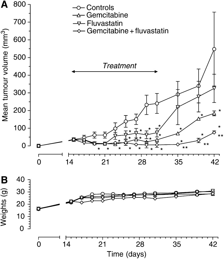
The new combination between the nucleoside analogue gemcitabine and the cholesterol-lowering drug fluvastatin was investigated in vitro and in vivo on the human pancreatic tumour cell line MIAPaCa-2. The present study demonstrates that fluvastatin inhibits proliferation, induces apoptosis in pancreatic cancer cells harbouring a p21ras mutation at codon 12 and synergistically potentiates the cytotoxic effect of gemcitabine. The pharmacologic activities of fluvastatin are prevented by administration of mevalonic acid, suggesting that the shown inhibition of geranyl-geranylation and farnesylation of cellular proteins, including p21rhoA and p21ras, plays a major role in its anticancer effect. Fluvastatin treatment also indirectly inhibits the phosphorylation of p42ERK2/mitogen-activated protein kinase, the cellular effector of ras and other signal transduction peptides. Moreover, fluvastatin administration significantly increases the expression of the deoxycytidine kinase, the enzyme required for the activation of gemcitabine, and simultaneously reduces the 5'-nucleotidase, responsible for deactivation of gemcitabine, suggesting a possible additional role of these enzymes in the enhanced cytotoxic activity of gemcitabine. Finally, a significant in vivo antitumour effect on MIAPaCa-2 xenografts was observed with the simultaneous combination of fluvastatin and gemcitabine, resulting in an almost complete suppression and a marked delay in relapse of tumour growth. In conclusion, the combination of fluvastatin and gemcitabine is an effective cytotoxic, proapoptotic treatment in vitro and in vivo against MIAPaCa-2 cells by a mechanism of action mediated, at least in part, by the inhibition of p21ras and rhoA prenylation. The obtained experimental findings might constitute the basis for a novel translational research in humans.


Figures

Figure 1
Figure 1
Dot-blot hybridisation analysis of point mutations (GGT → TGT) at codon 12 of the K-ras oncogene in MIAPaCa-2 and lung tumour; normal cornea (clone cell line) shows wild-type K-ras.
Figure 2
Figure 2
(A) Effect of fluvastatin, gemcitabine and their combination with mevalonic acid 100 μM on k-ras-mutated MIAPaCa-2 cell proliferation; (B) effect of fluvastatin and its combination with mevalonic acid 100 μM on wild-type k-ras COLO320-DM cell proliferation. Symbols and bars, mean values±s.e., respectively; *P<0.05 vs fluvastatin only.
Figure 3
Figure 3
Microscopic pictures of control MIAPaCa-2 cells (A) and cells treated with fluvastatin 2 μM (B) or gemcitabine 20 nM (C). Original magnification × 100.
Figure 4
Figure 4
Isobologram analysis of MIAPaCa-2 cell growth inhibition by (A) gemcitabine and fluvastatin simultaneously and sequentially, and (B) gemcitabine and PD098059 simultaneously. The IC75 values of each drug are plotted on the axes; the solid line represents the addictive effect, while the points representing the concentrations of gemcitabine and fluvastatin or PD098059 resulting in 75% growth inhibition of the combination are reported on the left of the connecting line, indicating synergism.
Figure 5
Figure 5
Deoxycytidine and 5′-NT expression in fluvastatin-treated MIAPaCa-2 cancer cells. Columns, mean values obtained from three independent experiments; bars, ±s.e.; *, statistically different from control cells (P<0.05).
Figure 6
Figure 6
(A) Gel electrophoresis of DNA extracted from fluvastatin- and gemcitabine-treated k-ras-mutated MIAPaCa-2 cells (upper). Image analysis of apoptotic DNA from cells exposed to fluvastatin, gemcitabine and their combination (lower); (B) gel electrophoresis of DNA extracted from 72 h fluvastatin-treated wild-type k-ras COLO320-DM cells. C, control; St, standard ladder.
Figure 7
Figure 7
In vitro effect of fluvastatin and gemcitabine on cell cycle distribution of MIAPaCa-2 cells. Percent values of cells in different phases of the cell cycle are given in each panel. The subdiploid peak in DNA histograms from treated cells indicates the occurrence of apoptosis (upper). The amount of subdiploid, apoptotic cells raises as the concentrations of the drugs in the culture media increase (lower). Columns and bars, mean values±s.e., respectively. *P<0.05 vs controls.
Figure 8
Figure 8
(A) Immunoblotting (upper) and image analysis (lower) of p21rhoA from total cellular lysates of MIAPaCa-2 cells treated with fluvastatin and gemcitabine. Columns and bars, mean values±s.e., respectively. MVA, mevalonic acid, G, geranyl-geranylated, N-G, nongeranyl-geranylated p21rhoA; *P<0.05 vs controls. (B) Immunoblotting (upper) and image analysis (lower) of p21ras from total cellular lysates of MIAPaCa-2 cells treated with fluvastatin and gemcitabine. Columns and bars, mean values±s.e., respectively. MVA, mevalonic acid, F, farnesylated, N-F, nonfarnesylated p21ras; *P<0.05 vs controls. (C) Immunoblotting (upper) and image analysis (lower) of p42MAPK/ERK2 in MIAPaCa-2 cells treated with fluvastatin and gemcitabine. Columns and bars, mean values±s.e., respectively. MVA, mevalonic acid, P, phosphorylated, N-P, nonphosphorylated p42MAPK/ERK2; *P<0.05 vs controls.
Figure 9
Figure 9
Immunohistochemical localisation (dark staining) of p21rhoA in MIAPaCa-2 cells control (A) and treated with fluvastatin (B), and p21ras in control cells (C) and treated with fluvastatin (D). Original magnification × 200.
Figure 10
Figure 10
(A) Chemotherapeutic effect of gemcitabine 120 mg kg−1 i.p. four times at 3-day intervals and fluvastatin 30 mg kg−1 i.p. every 2 days alone or in combination on MIAPaCa-2 tumours xenotransplanted in CD nu/nu mice. *P<0.05 with respect to controls; **P<0.05 vs gemcitabine and fluvastatin alone. Symbols and bars, mean±s.e. (B) Body weight of MIAPaCa-2 tumour-bearing control mice and mice treated with gemcitabine and fluvastatin alone or in combination. No changes or decline in body weight were noted. Symbols and bars, mean±s.e.

References

    1. Abbruzzese JL (2002a) New applications of gemcitabine and future directions in the management of pancreatic cancer. Cancer 95: 941–945 - PubMed
    1. Abbruzzese JL (2002b) Past and present treatment of pancreatic adenocarcinoma: chemotherapy as a standard treatment modality. Semin Oncol 29: 2–8 - PubMed
    1. Agarwal B, Bhendwal S, Halmos B, Moss SF, Ramey WG, Holt PR (1999) Lovastatin augments apoptosis induced by chemotherapeutic agents in colon cancer cells. Clin Cancer Res 5: 2223–2229 - PubMed
    1. Barton-Burke M (1999) Gemcitabine: a pharmacologic and clinical overview. Cancer Nurs 22: 176–183 - PubMed
    1. Bergman AM, Ruiz vHV, Veerman G, Kuiper CM, Peters GJ (1996) Synergistic interaction between cisplatin and gemcitabine in vitro. Clin Cancer Res 2: 521–530 - PubMed

MeSH terms