

Insular and gustatory inputs to the caudal ventral striatum in primates
- PMID: 16052493
- PMCID: PMC2474655
- DOI: 10.1002/cne.20660
Insular and gustatory inputs to the caudal ventral striatum in primates
Abstract
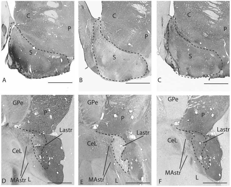
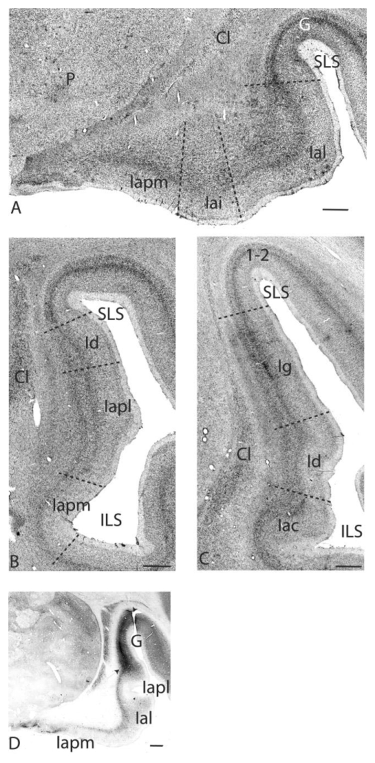
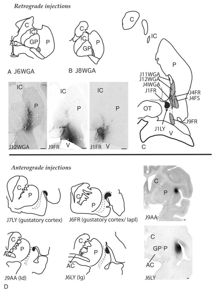
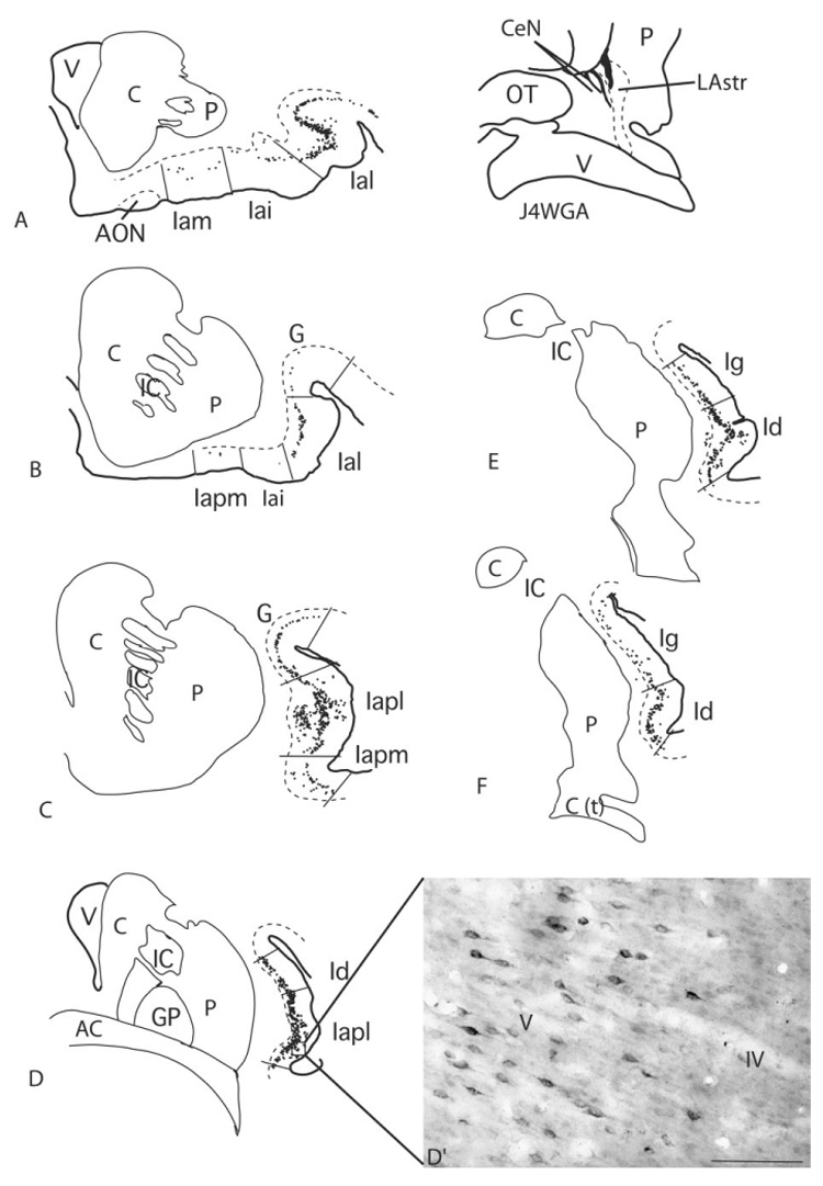
The ventral striatum mediates goal-directed behaviors based, in part, on inputs from the amygdala. However, striatal areas caudal to the ventral striatum also receive inputs from the amygdala. In primates, the amygdala projects to the central ventral putamen, lateral amygdalostriatal area, and caudal ventral putamen, suggesting that these regions are also "limbic-related." The anterior insula, which integrates sensory and amygdaloid inputs, projects to the classic ventral striatum. We used retrograde and anterograde tract tracing techniques to determine the extent to which specific subdivisions of the insula influence the caudal ventral striatum in the primate. The anterior (agranular and rostral dysgranular) insula has significant inputs to caudal ventral striatal regions that receive projections from the amygdala. In contrast, the posterior (granular) insula has sparse projections. Within the agranular insula, the posteromedial agranular (Iapm), lateral agranular (Ial), and posterolateral agranular (Iapl) subdivisions have the strongest inputs. These subdivisions mediate olfactory, gustatory, and visceral information processing (Carmichael and Price JL [1996b] J. Comp. Neurol. 363:642-640). In contrast, the intermediate agranular subdivision (Iai) is relatively devoid of visceral/gustatory inputs and has few inputs. In summary, caudal ventral striatal areas that receive amygdaloid inputs also receive significant innervation by agranular and dysgranular insula subdivisions that are themselves connected with the amygdala. Within this projection, the Ial, Iapm, and Iapl make the strongest contribution, suggesting that highly processed visceral/autonomic information, taste, and olfaction influence behavioral responses mediated by the caudal ventral striatum.
(c) 2005 Wiley-Liss, Inc.
Figures


Similar articles
-
Amygdaloid inputs define a caudal component of the ventral striatum in primates.J Comp Neurol. 2004 Aug 30;476(4):330-47. doi: 10.1002/cne.20228. J Comp Neurol. 2004. PMID: 15282709 Free PMC article.
-
Amygdaloid projections to ventromedial striatal subterritories in the primate.Neuroscience. 2002;110(2):257-75. doi: 10.1016/s0306-4522(01)00546-2. Neuroscience. 2002. PMID: 11958868
-
Insular cortical projections to functional regions of the striatum correlate with cortical cytoarchitectonic organization in the primate.J Neurosci. 1997 Dec 15;17(24):9686-705. doi: 10.1523/JNEUROSCI.17-24-09686.1997. J Neurosci. 1997. PMID: 9391023 Free PMC article.
-
Amygdala projections to central amygdaloid nucleus subdivisions and transition zones in the primate.Neuroscience. 2009 Mar 17;159(2):819-41. doi: 10.1016/j.neuroscience.2009.01.013. Neuroscience. 2009. PMID: 19272304 Free PMC article.
-
Efferent connections of the substantia innominata in the rat.J Comp Neurol. 1988 Nov 15;277(3):347-64. doi: 10.1002/cne.902770303. J Comp Neurol. 1988. PMID: 2461973
Cited by
-
Synchrony of anterior cingulate cortex and insular-striatal activation predicts ambiguity aversion in individuals with low impulsivity.Cereb Cortex. 2014 May;24(5):1397-408. doi: 10.1093/cercor/bht008. Epub 2013 Jan 24. Cereb Cortex. 2014. PMID: 23355606 Free PMC article.
-
Pay attention to the basal ganglia: a volumetric study in early dementia with Lewy bodies.Alzheimers Res Ther. 2019 Dec 21;11(1):108. doi: 10.1186/s13195-019-0568-y. Alzheimers Res Ther. 2019. PMID: 31864422 Free PMC article.
-
The Organization of the Primate Insular Cortex.Front Neuroanat. 2019 May 8;13:43. doi: 10.3389/fnana.2019.00043. eCollection 2019. Front Neuroanat. 2019. PMID: 31133822 Free PMC article. Review.
-
Understanding the neural circuitry of appetitive regulation in eating disorders.Biol Psychiatry. 2011 Oct 15;70(8):704-705. doi: 10.1016/j.biopsych.2011.08.018. Biol Psychiatry. 2011. PMID: 21967986 Free PMC article. No abstract available.
-
Maintenance and Representation of Mind Wandering during Resting-State fMRI.Sci Rep. 2017 Jan 12;7:40722. doi: 10.1038/srep40722. Sci Rep. 2017. PMID: 28079189 Free PMC article.
References
-
- Aggleton JP, Burton MJ, Passingham RE. Cortical and subcortical afferents to the amygdala of the rhesus monkey (Macaca mulatta) Brain Res. 1980;190:347–368. - PubMed
-
- Alexander GE, DeLong MR. Microstimulation of the primate neostriatum. II. Somatotopic organization of striatal microexcitable zones and their relation to neuronal response properties. J Neurophysiol. 1985;53:1417–1430. - PubMed
-
- Amaral DG, Price JL. Amygdalo-cortical projections in the monkey (Macaca fascicularis) J Comp Neurol. 1984;230:465–496. - PubMed
-
- Anderson AK, Christoff K, Stappen I, Panitz D, Ghahremani DG, Glover G, Gabrieli JD, Sobel N. Dissociated neural representations of intensity and valence in human olfaction.[see comment] Nat Neurosci. 2003;6:196–202. - PubMed
-
- Aosaki T, Kimura M, Graybiel AM. Temporal and spatial characteristics of tonically active neurons of the primate’s striatum. J Neurophysiol. 1995;73:1234–1252. - PubMed
Publication types
MeSH terms
Substances
Grants and funding
LinkOut - more resources
Full Text Sources
Miscellaneous
