

Antibody to eosinophil cationic protein suppresses dextran sulfate sodium-induced colitis in rats
- PMID: 16052679
- PMCID: PMC4398699
- DOI: 10.3748/wjg.v11.i29.4505
Antibody to eosinophil cationic protein suppresses dextran sulfate sodium-induced colitis in rats
Abstract
Aim: To produce an antibody against rat eosinophil cationic protein (ECP) and to examine the effects of the antibody in rats with dextran sulfate sodium (DSS)-induced colitis.
Methods: An antibody was raised against rat ECP. Rats were treated with 3% DSS in drinking water for 7 d and received the antibody or normal serum. The colons were examined histologically and correlated with clinical symptoms. Immunohistochemistry and Western blot analysis were estimated as a grade of inflammation.
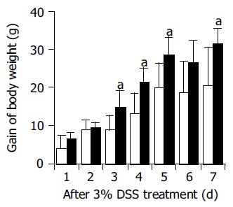
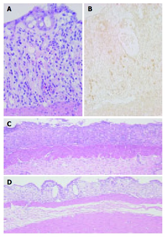
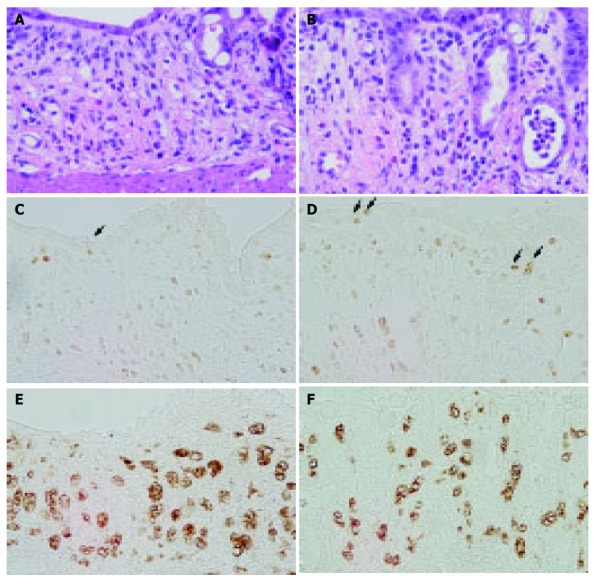
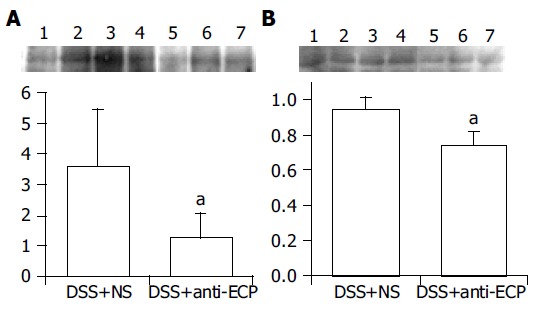
Results: The ECP antibody stained the activated eosinophils around the injured crypts in the colonic mucosa. Antibody treatment reduced the severity of colonic ulceration and acute clinical symptoms (diarrhea and/or blood-stained stool). Body weight gain was significantly greater and the colon length was significantly longer in anti-ECP-treated rats than in normal serum-treated rats. Expression of ECP in activated eosinophils was associated with the presence of erosions and inflammation. The number of Ki-67-positive cells in the regenerated surface epithelium increased in anti-ECP-treated rats compared with normal serum-treated rats. Western blot analysis revealed reduced expression of macrophage migration inhibitory factor (MIF) in anti-ECP-treated rats.
Conclusion: Our results indicate that treatment with ECP antibody, improved DSS-induced colitis in rats, possibly by increasing the regenerative activity of the colonic epithelium and downregulation of the immune response, and suggest that anti-ECP may promote intestinal wound healing in patients with ulcerative colitis (UC).
Figures
Similar articles
-
Prophylactic role of curcumin in dextran sulfate sodium (DSS)-induced ulcerative colitis murine model.Food Chem Toxicol. 2009 Jun;47(6):1311-7. doi: 10.1016/j.fct.2009.03.003. Epub 2009 Mar 12. Food Chem Toxicol. 2009. PMID: 19285535
-
Effect of toll-like receptor 3 agonist poly I:C on intestinal mucosa and epithelial barrier function in mouse models of acute colitis.World J Gastroenterol. 2017 Feb 14;23(6):999-1009. doi: 10.3748/wjg.v23.i6.999. World J Gastroenterol. 2017. PMID: 28246473 Free PMC article.
-
Jianpi Qingchang decoction alleviates ulcerative colitis by inhibiting nuclear factor-κB activation.World J Gastroenterol. 2017 Feb 21;23(7):1180-1188. doi: 10.3748/wjg.v23.i7.1180. World J Gastroenterol. 2017. PMID: 28275298 Free PMC article.
-
Protective effect of lactulose on dextran sulfate sodium-induced colonic inflammation in rats.Dig Dis Sci. 2004 Sep;49(9):1466-72. doi: 10.1023/b:ddas.0000042248.48819.ad. Dig Dis Sci. 2004. PMID: 15481321
-
Adenovirus-mediated hepatocarcinoma-intestine-pancreas/pancreatitis-associated protein suppresses dextran sulfate sodium-induced acute ulcerative colitis in rats.Inflamm Bowel Dis. 2012 Oct;18(10):1950-60. doi: 10.1002/ibd.22887. Epub 2012 Mar 14. Inflamm Bowel Dis. 2012. PMID: 22419609
Cited by
-
CCR3 Blockade Attenuates Eosinophilic Ileitis and Associated Remodeling.Am J Pathol. 2011 Nov;179(5):2302-14. doi: 10.1016/j.ajpath.2011.07.039. Epub 2011 Sep 23. Am J Pathol. 2011. PMID: 21945903 Free PMC article.
-
The effect of calendula officinalis in therapy of acetic Acid induced ulcerative colitis in dog as an animal model.Iran Red Crescent Med J. 2011 Dec;13(12):884-90. Epub 2011 Dec 1. Iran Red Crescent Med J. 2011. PMID: 22737434 Free PMC article.
-
Antieosinophil Antibodies Alone or in Combination with Antineutrophil Cytoplasmic Antibodies (ANCA) Detected in Different Autoimmune Conditions.J Immunol Res. 2023 Apr 26;2023:5980287. doi: 10.1155/2023/5980287. eCollection 2023. J Immunol Res. 2023. PMID: 37153639 Free PMC article.
-
CD34 is required for infiltration of eosinophils into the colon and pathology associated with DSS-induced ulcerative colitis.Am J Pathol. 2010 Sep;177(3):1244-54. doi: 10.2353/ajpath.2010.100191. Epub 2010 Aug 9. Am J Pathol. 2010. PMID: 20696776 Free PMC article.
-
Clinical value of serum eosinophilic cationic protein assessment in children with inflammatory bowel disease.Arch Med Sci. 2014 Dec 22;10(6):1142-6. doi: 10.5114/aoms.2013.34415. Epub 2013 Apr 9. Arch Med Sci. 2014. PMID: 25624851 Free PMC article.
References
-
- Dvorak AM. Ultrastructural evidence for release of major basic protein-containing crystalline cores of eosinophil granules in vivo: cytotoxic potential in Crohn's disease. J Immunol. 1980;125:460–462. - PubMed
-
- Sarin SK, Malhotra V, Sen Gupta S, Karol A, Gaur SK, Anand BS. Significance of eosinophil and mast cell counts in rectal mucosa in ulcerative colitis. A prospective controlled study. Dig Dis Sci. 1987;32:363–367. - PubMed
-
- Walsh RE, Gaginella TS. The eosinophil in inflammatory bowel disease. Scand J Gastroenterol. 1991;26:1217–1224. - PubMed
-
- Desreumaux P, Nutten S, Colombel JF. Activated eosinophils in inflammatory bowel disease: do they matter? Am J Gastroenterol. 1999;94:3396–3398. - PubMed
-
- Nishitani H, Okabayashi M, Satomi M, Shimoyama T, Dohi Y. Infiltration of peroxidase-producing eosinophils into the lamina propria of patients with ulcerative colitis. J Gastroenterol. 1998;33:189–195. - PubMed
Publication types
MeSH terms
Substances
LinkOut - more resources
Full Text Sources
Medical
Miscellaneous
