Review
. 2005 Jul;96(7):379-86.
doi: 10.1111/j.1349-7006.2005.00062.x.

Regulation of cancer cell motility through actin reorganization

Affiliations
Review

Regulation of cancer cell motility through actin reorganization

Daisuke Yamazaki et al. Cancer Sci. 2005 Jul.

Abstract

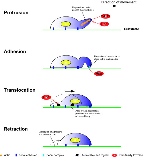
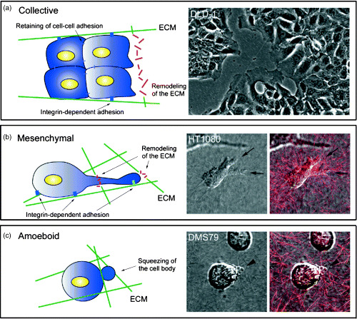
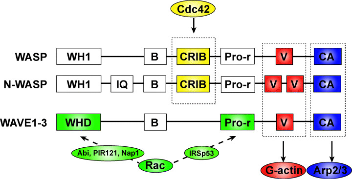
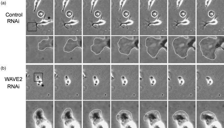
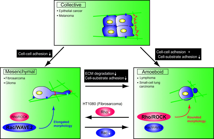
Cell migration is a critical step in tumor invasion and metastasis, and regulation of this process will lead to appropriate therapies for treating cancer. Cancer cells migrate in various ways, according to cell type and degree of differentiation. The different types of cell migration are regulated by different mechanisms. Reorganization of the actin cytoskeleton is the primary mechanism of cell motility and is essential for most types of cell migration. Actin reorganization is regulated by Rho family small GTPases such as Rho, Rac, and Cdc42. These small GTPases transmit extracellular chemotactic signals to downstream effectors. Of these downstream effectors, Wiskott-Aldrich syndrome protein (WASP) family proteins are key regulators of cell migration. Activated WASP family proteins induce the formation of protrusive membrane structures involved in cell migration and degradation of the extracellular matrix. Inhibition of Rho family small GTPase signaling suppresses the migration and invasion of cancer cells. Thus, control of cell migration via the actin cytoskeleton provides the possibility of regulating cancer cell invasion and metastasis.


Figures

Figure 1
Figure 1
Cell migration on 2‐D substrates. Cell migration consists of the following four successive processes. Protrusion: when cells are polarized by extracellular stimuli, de novo actin polymerization occurs at the leading edge; polymerized actin filaments induce the formation of membrane protrusions such as filopodia and lamellipodia. Adhesion: protruded membranes contact the substrate and form novel integrin‐dependent cell‐substrate adhesions; newly formed adhesion complexes are stabilized and mature into focal adhesions. Translocation: the nucleus and cell body are translocated by acto‐myosin contractile forces. Retraction: cell‐substrate adhesive structures at the trailing edge are disassembled, and the trailing edge retracts.
Figure 2
Figure 2
Modes of migration in cancer cells. Migration of cancer cells involves the following three processes. (a) Collective cell migration. Cells move as sheet‐like structures, maintaining cell–cell adhesions. The image shows migration of DLD‐1 cells on a 2‐D substrate. (b) Mesenchymal migration. In 3‐D matrices, cells form membrane protrusions at the leading edge and adhere to the substrate in an integrin‐dependent manner. By releasing extracellular matrix (ECM)‐degrading enzymes, cells remodel the ECM to form a path. The images show the migration of HT1080 cells in collagen gels. Arrows indicate membrane protrusions at the leading edge. The red signal indicates the backscatter of collagen fibers. (c) Amoeboid migration. Cell‐substrate adhesion is weak and is independent of integrin function. Cells move by acto‐myosin contractile force and move within the ECM by squeezing the cell body. The images show the migration of DMS79 cells in collagen gels. Arrowheads indicate membrane blebbing. The red signal indicates the backscatter of collagen fibers.
Figure 3
Figure 3
The Wiskott–Aldrich syndrome protein (WASP) family proteins. WASP family proteins consist of multiple functional domains. WASP and neural WASP (N‐WASP) are activated by direct binding to Cdc42 via the CRIB domain, whereas WAVE (WASP‐family verprolin‐homologous proteins) bind to Rac indirectly via various molecules. The C‐terminal verprolin‐homology, cofilin‐like, and acidic (VCA) domains are important for the induction of actin polymerization. The V and CA domains bind actin monomer (G‐actin) and the Arp2/3 complex, respectively. WASP family proteins activated by Rac and Cdc42 induce Arp2/3 complex‐dependent actin polymerization via the VCA domain. B, basic; CA, cofilin‐like and acidic; CRIB, Cdc42 and Rac interactive binding; IQ, IQ motif; Pro‐r, Proline‐rich; V, verprolin homology; WH1, WASP homology 1; WHD, WAVE homology domain.
Figure 4
Figure 4
The WAVE2 (Wiskott–Aldrich syndrome protein family verprolin‐homologous protein 2) is required for cancer cell migration in 3‐D matrices. ( 24 ) Time‐lapse phase‐contrast images of RNA‐interference (RNAi)‐treated B16F10 cells in extracellular matrix‐containing gels. Asterisks represent transfected cells. Repression of WAVE2 expression by RNAi methods reduces membrane protrusions at the leading edge, which are important for migration and invasion of melanoma cells. Magnified images of boxed areas are shown in the lower panels.
Figure 5
Figure 5
Transition of migration in cancer cells. When cell–cell adhesions are lost by inhibition of cadherin function, cancer cells that undergo collective migration begin to move separately (mesenchymal migration). When both cell–cell and cell–substrate adhesion are inhibited simultaneously, collective migration changes to amoeboid. Inhibition of extracellular matrix degradation and integrin‐dependent adhesion cause mesenchymal‐amoeboid transition (MAT). In HT1080 fibrosarcoma cells, which show both mesenchymal and amoeboid types of cell migration, inhibition of Rac signaling induces MAT. In contrast, inhibition of Rho/Rho‐kinase (ROCK) signaling induces an elongated morphology.

References

    1. Ridley AJ, Schwartz MA, Burridge K et al. Cell migration: integrating signals from front to back. Science 2003; 302: 1704–9. - PubMed
    1. Pollard TD, Borisy GG. Cellular motility driven by assembly and disassembly of actin filaments. Cell 2003; 112: 453–65. - PubMed
    1. Condeelis J, Segall JE. Intravital imaging of cell movement in tumours. Nat Rev Cancer 2003; 3: 921–30. - PubMed
    1. Kundra V, Escobedo JA, Kazlauskas A et al. Regulation of chemotaxis by the platelet‐derived growth factor receptor‐beta. Nature 1994; 367: 474–6. - PubMed
    1. Huttenlocher A, Sandborg RR, Horwitz AF. Adhesion in cell migration. Curr Opin Cell Biol 1995; 7: 697–706. - PubMed

MeSH terms