Comparative Study
. 2005 Jul;59(7):861-7.
doi: 10.1366/0003702054411616.

Simple Raman instrument for in vivo detection of macular pigments

Affiliations
Comparative Study

Simple Raman instrument for in vivo detection of macular pigments

Igor V Ermakov et al. Appl Spectrosc. 2005 Jul.

Abstract

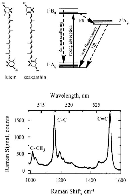
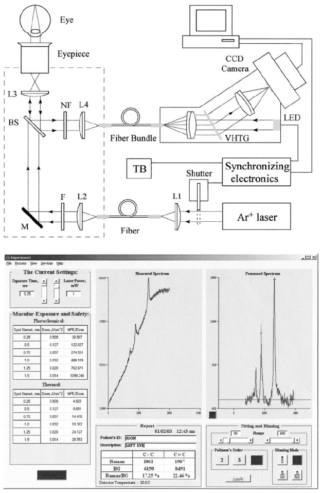
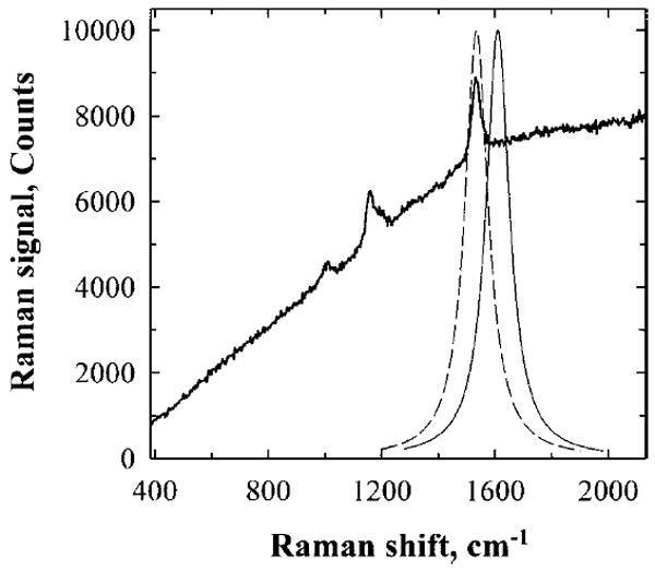
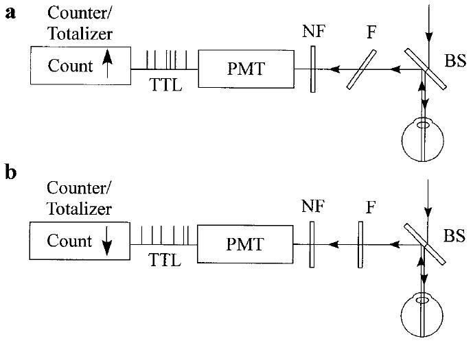
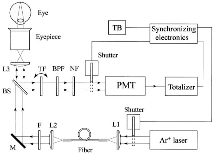
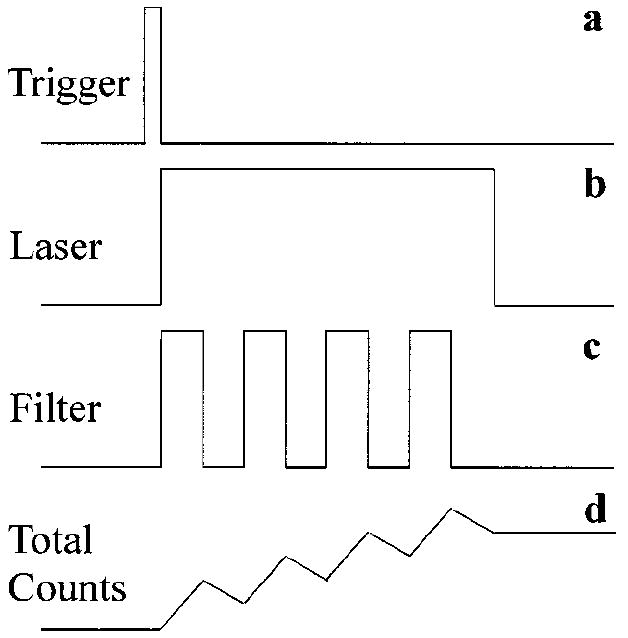
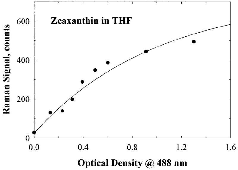
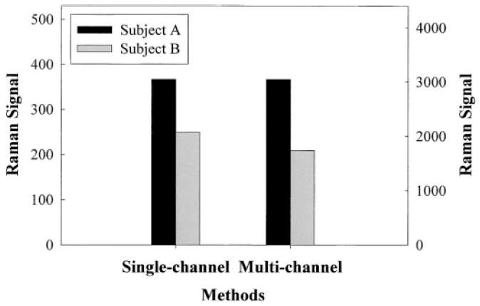
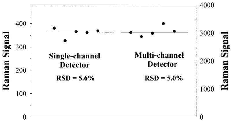
Raman spectroscopy holds promise as a novel noninvasive technology for the quantification of the macular pigments (MP) lutein and zeaxanthin. These compounds, which are members of the carotenoid family, are thought to prevent or delay the onset of age-related macular degeneration, the leading cause of irreversible blindness in the elderly. It is highly likely that they achieve this protection through their function as optical filters and/or antioxidants. Using resonant excitation in the visible region, we measure and quantify the Raman signals that originate from the carbon double bond (C=C) stretch vibrations of the pi-conjugated molecule backbone. In this manuscript we describe the construction and performance of a novel compact MP Raman instrument utilizing dielectric angle-tuned band-pass filters for wavelength selection and a single-channel photo-multiplier for the detection of MP Raman responses. MP concentration measurements are fast and accurate, as seen in our experiments with model eyes and living human eyes. The ease and rapidity of Raman MP measurements, the simplicity of the instrumentation, the high accuracy of the measurements, and the lack of significant systematic errors should make this technology attractive for widespread clinical research.


Figures

Fig. 1
Fig. 1
(Left panel) Chemical structure, (right panel) energy level diagram, and (lower panel) resonance Raman spectrum of the macular carotenoids lutein and zeaxanthin. The three most prominent carotenoid Raman peaks occur at 1008, 1159, and 1524 cm−1, as shown for a solution of lutein in tetrahydrofuran (THF), and correspond, respectively, to rocking motions of methyl components (C–CH3) and stretch vibrations of the carbon–carbon single (C–C) and double bonds (C=C). The side peak at 1030 cm−1 is an artifact and corresponds to THF. Laser excitation was at 488 nm. A fluorescence background was subtracted.
Fig. 2
Fig. 2
(Upper panel) Optical diagram of the dispersive multi-channel MP Raman detector and (lower panel) MP Raman spectra obtained with it.
Fig. 3
Fig. 3
Operational principles of single-channel Raman detection. The intensity of the carotenoid C=C Raman peak can be measured with a tunable narrow band pass filter and single-channel light detector. When the transmission band of the filter is tuned to the C=C peak (dashed curve), both Raman signal and accompanying background fluorescence are measured. When the filter’s transmission curve is slightly shifted from the Raman peak (solid curve), the detector registers only background fluorescence. The subtraction of the latter signal from the former one gives the C=C Raman signal intensity.
Fig. 4
Fig. 4
Technical implementation of single-channel MP Raman detection. Blue excitation laser light is routed into the human eye via a dichroic beam splitter (BS). Backscattered Raman light is reflected toward the registration module. A thin, angle-tunable dielectric band pass filter F, transmits, depending on its orientation, either (a) the sum of the retinal fluorescence and carotenoid Raman signal, or (b) retinol fluorescence only. The electronics of a photo multiplier tube (PMT) produces TTL pulses with each photon detection event. Note that the pulse counter/totalizer is synchronized with the angle-tuned filter: it counts UP whenever the filter transmits at the Raman wavelength, and it counts DOWN whenever pure retinal fluorescence is transmitted.
Fig. 5
Fig. 5
Optical diagram of the single-channel version of the MP Raman detector.
Fig. 6
Fig. 6
Timing diagram of single-channel measurements. A trigger button initiates measurements providing a trigger signal (a) followed by laser shutter activation (b). Laser light remains ON during several cycles of MP measurements. An angle-tunable filter changes its orientation once per cycle (c) and a pulse counter continues to add up or subtract pulse counts according to the filter position (d). Multiple measurements assure high signal-to-noise ratios.
Fig. 7
Fig. 7
Calibration curve obtained for single-channel MP Raman detection. It links the reading provided by the instrument with the carotenoid content in a human macula. The curve was obtained using a model eye attached to the instrument. The model eye discussed in the text contains zeaxanthin dissolved in methanol. The sampling volume, 1 mL, corresponds to a 1-mm-diameter spot at the macula and contains a carotenoid concentration typical for a human macula.
Fig. 8
Fig. 8
Comparison of single- and multi-channel instruments regarding MP Raman responses of two volunteer subjects, A and B. The Raman signal strength at left corresponds to single-channel detection and the scale at right to multi-channel detection, respectively. The signal strengths obtained with the two instruments for subject A are shown at the same level to facilitate comparison of the responses for subject B.
Fig. 9
Fig. 9
Test-retest repeatability and average values of MP measurements for a subject obtained with single-channel and multi-channel Raman detection. The Raman signal strength at left corresponds to single-channel detection and the scale at right to multi-channel detection, respectively.

Comment in

Similar articles

Cited by

References

    1. Schalch W, Dayhaw-Barker P, Barker FM. The carotenoids of the human retina. In: Taylor A, editor. Nutritional and Environmental Influences on the Eye. CRC; Boca Raton, FL: 1999. pp. 215–250.
    1. Seddon JM, Ajani UA, Sperduto RD, Hiller R, Blair N, Burton TC, Farber MD, Gragoudas ES, Haller J, Miller DT, Yanuzzi LA, Willet W. J Am Med Assoc. 1994;272:1413. - PubMed
    1. Beatty S, Koh H-H, Henson D, Boulton M. Surv Ophthalmol. 2000;45:115. - PubMed
    1. Reading VM, Weale RA. J Am Optom Assoc. 1974;64:231.
    1. Beatty S, Murray IJ, Henson DB, Carden D, Koh H-H, Boulton ME. Invest Ophthalmol Visual Sci. 2001;42:439. - PubMed

Publication types

MeSH terms

Substances