. 2005 Jun;1(6):393-9.
doi: 10.1016/j.cmet.2005.05.003.

Mitochondrial dysfunction resulting from loss of cytochrome c impairs cellular oxygen sensing and hypoxic HIF-alpha activation

Affiliations

Mitochondrial dysfunction resulting from loss of cytochrome c impairs cellular oxygen sensing and hypoxic HIF-alpha activation

Kyle D Mansfield et al. Cell Metab. 2005 Jun.

Abstract

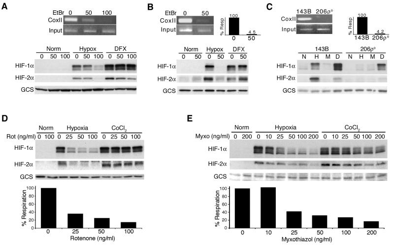
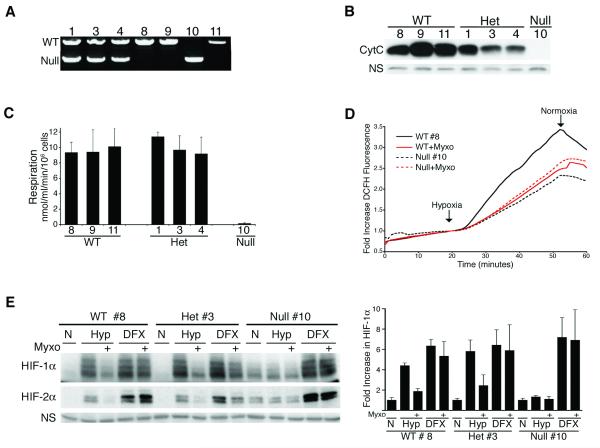
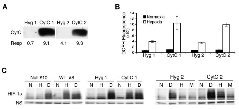
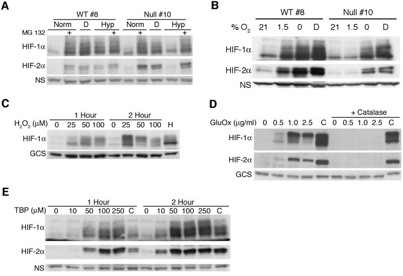
While cellular responses to low oxygen (O(2)) or hypoxia have been studied extensively, the precise identity of mammalian cellular O(2) sensors remains controversial. Using murine embryonic cells lacking cytochrome c, and therefore mitochondrial activity, we show that mitochondrial reactive oxygen species (mtROS) are essential for proper O(2) sensing and subsequent HIF-1 alpha and HIF-2 alpha stabilization at 1.5% O(2). In the absence of this signal, HIF-alpha subunits continue to be degraded. Furthermore, exogenous treatment with H(2)O(2) or severe O(2) deprivation is sufficient to stabilize HIF-alpha even in the absence of cytochrome c and functional mitochondria. These results provide genetic evidence indicating that mtROS act upstream of prolyl hydroxylases in regulating HIF-1 alpha and HIF-2 alpha in this O(2)-sensing pathway.


Figures

Figure 1
Figure 1
Functional mitochondria are required for hypoxic HIF-α stabilization. A) Hep3B cells treated with 0, 50 or 100 ng/ml EtBr for 3 weeks and mtDNA levels determined via COXII PCR. Hep3B control (0 and 50) or ρ° (100) cells exposed to 4 hours of normoxia (21% O2), hypoxia (1.5% O2) or DFX (100 μM) and analyzed for HIF-1α and HIF-2α via western blot with γ-glutamylcysteine synthetase (GCS) as a loading control. B) HEK293 cells treated for 2 weeks with 0 or 50 ng/ml EtBr analyzed for COXII mtDNA and respiration ((Resp, expressed as nmol O2 consumed/ml/min/106 cells). Control (0) or ρ° (50) cells exposed to 4 hours of normoxia, hypoxia or DFX. C) 143B and 206ρ° cells analyzed for COXII mtDNA and respiration. Cells exposed to normoxia (N), hypoxia (H), hypoxia plus 100 ng/ml myxothiazol (M), or DFX (D) for 4 hours. D, E) Hep3B cells treated with rotenone (D) or myxothiazol (E) and exposed to hypoxia or 100 μM CoCl2 for 4 hours. HIF-1α and HIF-2α levels determined and respiration measured to confirm mitochondrial inhibition.
Figure 2
Figure 2
Cytochrome c null embryonic cells are deficient in their hypoxic response. A) Genotype of individual cell lines determined via PCR. B) Cytochrome c protein expression determined via western blot. C) Mitochondrial respiration was measured. D) Embryonic cells mounted in a flow-through chamber and ROS production in response to 1% O2 was monitored with DCFH-DA. mtROS levels determined by treatment with 100 ng/ml myxothiazol. E) Embryonic cells exposed to normoxia (N), hypoxia (Hyp, 1.5% O2) or 100 μM DFX for 4 hours in the presence or absence of 100 ng/ml myxothiazol and HIF-α levels determined. Densitometry analysis of HIF-1α levels in 5 separate experiments normalized to normoxic controls.
Figure 3
Figure 3
Reintroduction of cytochrome c restores hypoxic response. A) An independent cytochrome c null cell line stably transfected with a control (Hyg), or cytochrome c expression vector (CytC) and assayed for cytochrome c protein levels and respiratory activity. B) ROS accumulation monitored with carboxy-H2 DCFDA and fluorescence determined (+/− S.E) after 4 hours of hypoxia. C) Cells exposed to 4 hours of normoxia (N), 100 μM DFX (D), or hypoxia in the absence (H) or presence (M) of 100 ng/ml myxothiazol and HIF-1α expression determined.
Figure 4
Figure 4
Proteosome inhibition, anoxia, or H2O2 is sufficient to stabilize HIF-α in cytochrome c null cells. A) Cytochrome c WT or null cells exposed to 4 hours of normoxia or hypoxia in the presence of 10 μM MG132 or 100 μM DFX and HIF-α levels determined. B) Embryonic cells exposed to 21%, 1.5%, and 0% O2, or 100 μM DFX for 3 hours. C) Hep3B cells treated with H2O2 or exposed to hypoxia (H) for 2 hours. D) Hep3B cells treated with glucose oxidase enzyme (Gluc Ox) or CoCl2 for 2 hours in the presence or absence of 100 units/ml catalase. E) Cytochrome c null cells treated with boluses of t-Butyl hydroperoxide (TBP) every 15 mins or a single dose of CoCl2 (100 μM) for 1 or 2 hours.

Similar articles

Cited by

References

    1. Agani FH, Pichiule P, Chavez J. Carlos, LaManna JC. Inhibitors of mitochondrial complex I attenuate the accumulation of hypoxia-inducible factor-1 during hypoxia in Hep3B cells. Comp Biochem Physiol A Mol Integr Physiol. 2002a;132:107–109. - PubMed
    1. Agani FH, Pichiule P, Chavez JC, LaManna JC. The role of mitochondria in the regulation of hypoxia-inducible factor 1 expression during hypoxia. J Biol Chem. 2000;275:35863–35867. - PubMed
    1. Agani FH, Puchowicz M, Chavez JC, Pichiule P, LaManna J. Role of nitric oxide in the regulation of HIF-1alpha expression during hypoxia. Am J Physiol Cell Physiol. 2002b;283:C178–186. - PubMed
    1. Barros MH, Netto LE, Kowaltowski AJ. H(2)O(2) generation in Saccharomyces cerevisiae respiratory pet mutants: effect of cytochrome c. Free Radic Biol Med. 2003;35:179–188. - PubMed
    1. BelAiba RS, Djordjevic T, Bonello S, Flugel D, Hess J, Kietzmann T, Gorlach A. Redox-sensitive regulation of the HIF pathway under non-hypoxic conditions in pulmonary artery smooth muscle cells. Biol Chem. 2004;385:249–257. - PubMed

Publication types

LinkOut - more resources