

Lower respiratory tract infection due to Capnocytophaga cynodegmi in a cat with pulmonary carcinoma
- PMID: 16055008
- PMCID: PMC10822334
- DOI: 10.1016/j.jfms.2004.11.002
Lower respiratory tract infection due to Capnocytophaga cynodegmi in a cat with pulmonary carcinoma
Abstract
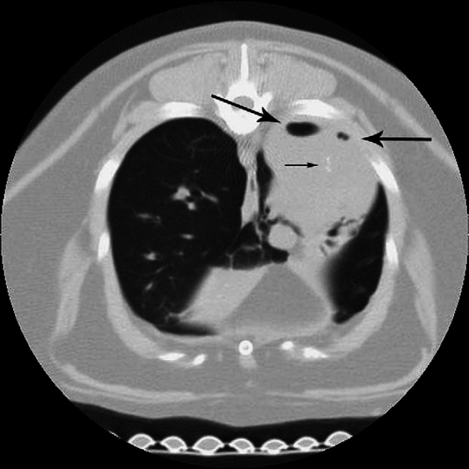
A 10-year-old male castrated domestic shorthair cat was evaluated for coughing and lethargy. Thoracic radiographs revealed a soft tissue lung mass and diffuse peribronchial infiltrates. Bronchoscopy was performed and Capnocytophaga cynodegmi was cultured from bilateral bronchoalveolar lavage samples. Clinical signs and bacterial colonization resolved following treatment with enrofloxacin. A lung lobectomy was performed to remove the lung mass, which was diagnosed as pulmonary carcinoma. C cynodegmi is most frequently isolated from localized wound and corneal infections in humans. Specialized growth characteristics of C cynodegmi may result in low sensitivity for bacterial culture. To the authors' knowledge, this case represents the first report of C cynodegmi infection in a veterinary patient and only the second case in human or veterinary medicine where the organism has been isolated from a bronchoalveolar lavage sample. Based on this report, Capnocytophaga species should be considered as potential opportunistic pathogens.
Figures

Similar articles
-
[The presence of Capnocytophaga canimorsus and Capnocytophaga cynodegmi in companion animals in the Netherlands].Tijdschr Diergeneeskd. 2011 Jul 1;136(7):490-2. Tijdschr Diergeneeskd. 2011. PMID: 21870573 Dutch.
-
Molecular characterization of Capnocytophaga canimorsus and other canine Capnocytophaga spp. and assessment by PCR of their frequencies in dogs.J Clin Microbiol. 2009 Oct;47(10):3218-25. doi: 10.1128/JCM.01246-09. Epub 2009 Jul 29. J Clin Microbiol. 2009. PMID: 19641058 Free PMC article.
-
Prevalence of Capnocytophaga canimorsus and Capnocytophaga cynodegmi in dogs and cats determined by using a newly established species-specific PCR.Vet Microbiol. 2010 Jul 29;144(1-2):172-6. doi: 10.1016/j.vetmic.2010.01.001. Epub 2010 Jan 18. Vet Microbiol. 2010. PMID: 20144514
-
Capnocytophaga canimorsus.Vet Microbiol. 2010 Jan 27;140(3-4):339-46. doi: 10.1016/j.vetmic.2009.01.040. Epub 2009 Feb 5. Vet Microbiol. 2010. PMID: 19268498 Review.
-
Lower respiratory tract infections in cats: reaching beyond empirical therapy.J Feline Med Surg. 2011 May;13(5):313-32. doi: 10.1016/j.jfms.2011.03.009. J Feline Med Surg. 2011. PMID: 21515220 Free PMC article. Review.
Cited by
-
Outcome and prognostic indicators in 20 cats with surgically treated primary lung tumors.J Feline Med Surg. 2014 Dec;16(12):979-84. doi: 10.1177/1098612X14530121. Epub 2014 Apr 7. J Feline Med Surg. 2014. PMID: 24710595 Free PMC article.
-
Draft Genome Sequences of Three Capnocytophaga cynodegmi Strains Isolated from the Oral Cavity of Healthy Dogs.Genome Announc. 2015 May 28;3(3):e00200-15. doi: 10.1128/genomeA.00200-15. Genome Announc. 2015. PMID: 26021913 Free PMC article.
-
Capnocytophaga cynodegmi in a rottweiler dog with severe bronchitis and foreign-body pneumonia.J Clin Microbiol. 2008 Dec;46(12):4099-103. doi: 10.1128/JCM.00173-08. Epub 2008 Aug 27. J Clin Microbiol. 2008. PMID: 18753348 Free PMC article.
-
Capnocytophaga cynodegmi peritonitis in a peritoneal dialysis patient.J Clin Microbiol. 2007 Nov;45(11):3844-6. doi: 10.1128/JCM.00761-07. Epub 2007 Sep 19. J Clin Microbiol. 2007. PMID: 17881542 Free PMC article.
-
Discovery and validation of potential bacterial biomarkers for lung cancer.Am J Cancer Res. 2015 Sep 15;5(10):3111-22. eCollection 2015. Am J Cancer Res. 2015. PMID: 26693063 Free PMC article.
References
-
- August J.R. Dysgonic fermenter-2 infections, Journal of the American Veterinary Medical Association 193, 1988, 1506–1508. - PubMed
-
- Blanche P., Bloch E., Sicard D. Capnocytophaga canimorsus in the oral flora of dogs and cats, Journal of Infection 36, 1998, 134. - PubMed
-
- Brenner D.J., Hollis D.G., Fanning G.R., Weaver R.E. Capnocytophaga canimorsus species nov. (formerly CDC group DF-2), a cause of septicemia following dog bite, and C cynodegmi sp. nov., a cause of localized wound infection following dog bite, Journal of Clinical Microbiology 27, 1989, 231–235. - PMC - PubMed
-
- Chambers G.W., Westblom T.U. Pleural infection caused by Capnocytophaga canimorsus, formerly CDC group DF-2, Clinical Infectious Disease 15, 1992, 325–326. - PubMed
Publication types
MeSH terms
LinkOut - more resources
Full Text Sources
Medical
Miscellaneous
