. 2005 Oct;16(10):4905-17.
doi: 10.1091/mbc.e05-01-0019. Epub 2005 Jul 29.

Nuclear aggresomes form by fusion of PML-associated aggregates

Affiliations

Nuclear aggresomes form by fusion of PML-associated aggregates

Lianwu Fu et al. Mol Biol Cell. 2005 Oct.

Abstract

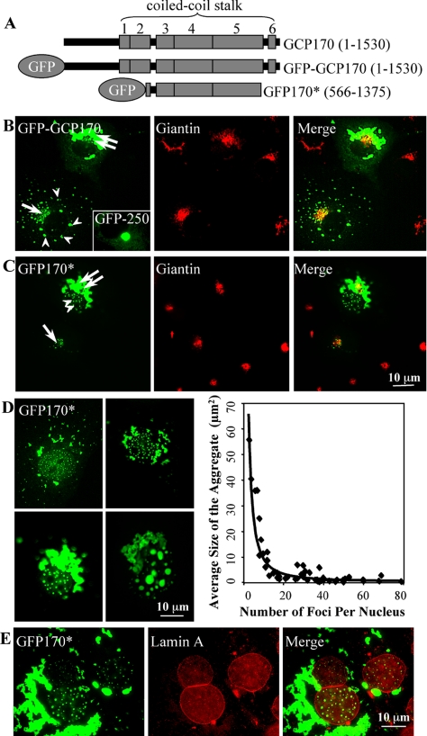
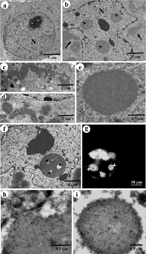
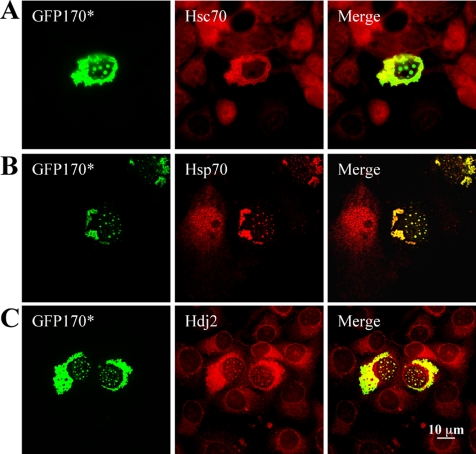
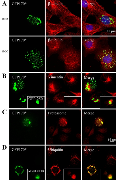
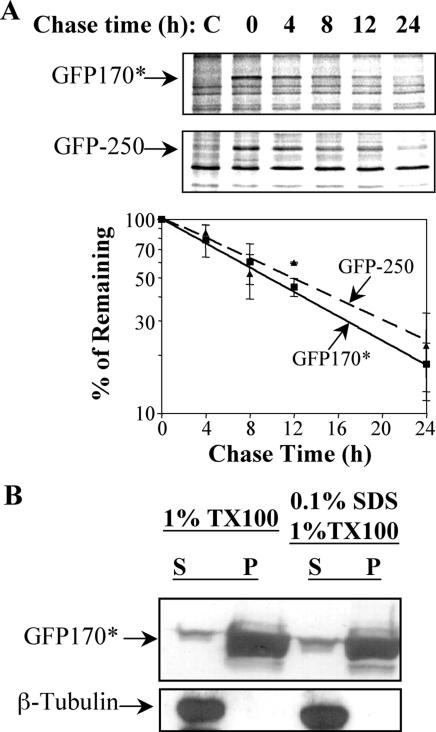
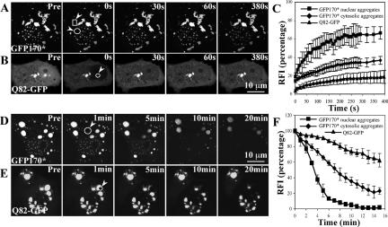
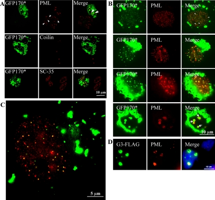
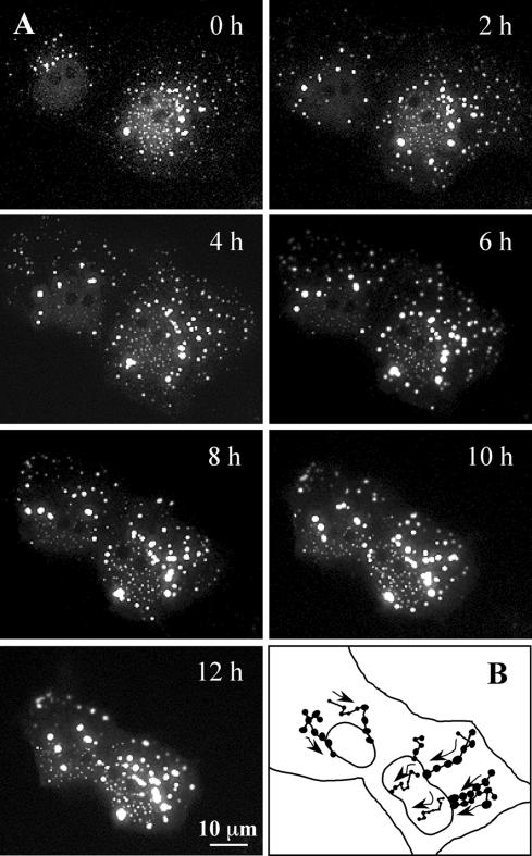
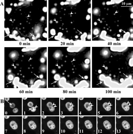
Nuclear aggregates formed by proteins containing expanded poly-glutamine (poly-Q) tracts have been linked to the pathogenesis of poly-Q neurodegenerative diseases. Here, we show that a protein (GFP170*) lacking poly-Q tracts forms nuclear aggregates that share characteristics of poly-Q aggregates. GFP170* aggregates recruit cellular chaperones and proteasomes, and alter the organization of nuclear domains containing the promyelocytic leukemia (PML) protein. These results suggest that the formation of nuclear aggregates and their effects on nuclear architecture are not specific to poly-Q proteins. Using GFP170* as a model substrate, we explored the mechanistic details of nuclear aggregate formation. Fluorescence recovery after photobleaching and fluorescence loss in photobleaching analyses show that GFP170* molecules exchange rapidly between aggregates and a soluble pool of GFP170*, indicating that the aggregates are dynamic accumulations of GFP170*. The formation of cytoplasmic and nuclear GFP170* aggregates is microtubule-dependent. We show that within the nucleus, GFP170* initially deposits in small aggregates at or adjacent to PML bodies. Time-lapse imaging of live cells shows that small aggregates move toward each other and fuse to form larger aggregates. The coalescence of the aggregates is accompanied by spatial rearrangements of the PML bodies. Significantly, we find that the larger nuclear aggregates have complex internal substructures that reposition extensively during fusion of the aggregates. These studies suggest that nuclear aggregates may be viewed as dynamic multidomain inclusions that continuously remodel their components.


Figures

Figure 1.
Figure 1.
GFP170* deposits within the cytoplasm and in the nucleus. (A) Schematic diagram of full-length GCP170(1-1530), GFP-GCP170, and GFP170*(566-1375). The full-length GCP170 contains an amino-terminal head domain followed by a coiled-coil stalk of six-coiled coils (shaded boxes). GFP170* contains GFP fused to an internal segment (amino acid 566-1375 of GCP170). (B) COS-7 cells were transfected with a GFP-tagged full-length GCP170 or GFP-250 construct. After 48 h, cells were processed for indirect immunofluorescence using antibody against the Golgi marker protein giantin. In a cell expressing low levels of GFP-GCP170 (bottom left), most of the molecules are targeted to the Golgi region and colocalize with giantin (arrow). In addition, small peripheral aggregates are visible (arrowheads). In a cell expressing high levels of GFP-GCP170 (top right), the GFP-GCP170 forms large aggregates that surround the Golgi (double arrow). The GFP170* aggregates seem “ribbon-like” and are distinct from the spherical aggregates formed by GFP-250 (inset). (C) COS-7 cells were transfected with GFP170*. After 48 h, cells were processed for indirect immunofluorescence using antibody against giantin. In a cell expressing low levels of GFP170* (bottom left), most of the molecules are targeted to the Golgi region, as demonstrated by colocalization with giantin (arrow). In a cell expressing high levels of GFP170* (top right), GFP170* forms large aggregates that surround the Golgi (double arrow). In addition, GFP170* is detected in numerous nuclear foci (double arrowhead). (D) COS-7 cells were transfected with GFP170*. After 24-48 h, cells were processed for epifluorescence. Images of COS-7 cells containing different numbers of nuclear aggregates were selected. The average size of GFP170* nuclear foci was determined using IPLab software and plotted as a function of number of foci per nucleus. (E) COS-7 cells were transfected with GFP170*. After 24 h, cells were processed for immunofluorescence using anti-lamin A antibody. Spherical GFP170* foci are enclosed within the lamin A-defined space.
Figure 2.
Figure 2.
Ultrastructure of GFP170* aggregates. Nontransfected COS-7 cells (a) or COS-7 cells transfected with GFP170* (b-i) were processed for transmission electron microscopy (b-f), fluorescence (g), or immunogold labeling (h and i). (a) Nontransfected COS-7 cell lacks aggregates. (b) Transfected COS-7 cell contains cytoplasmic aggregates (arrows) and nuclear aggregates (arrowheads). (c and d) Cytoplasmic aggregates are ribbon-like and are surrounded by mitochondria. (e) Nuclear aggregates are either spherical or ovoid. (f) Nuclear aggregates contain internal electron-lucent spaces (arrowheads). (g) Internal GFP170* substructure within the nuclear aggregates is also visible by fluorescence (arrowheads). (h and i) The content of the cytoplasmic and nuclear aggregates was confirmed by immunogold labeling with anti-GFP antibodies followed by secondary antibodies conjugated to 12-nm gold particles (arrowheads). N, nucleus.
Figure 3.
Figure 3.
Recruitment of molecular chaperones to GFP170* aggregates. COS-7 cells were transfected with GFP170*. After 48 h, cells were processed for indirect immunofluorescence using antibodies against Hsc70 (A), Hsp70 (B), or Hdj2 (C). Chaperones are recruited to GFP170* aggregates.
Figure 4.
Figure 4.
Involvement of microtubules in the formation of GFP170* aggregates and the recruitment of vimentin and proteasomal components to GFP170* aggregates. (A) COS-7 cells were transfected with GFP170*. After 8 h, cells were left untreated (-noc) or supplemented with 1.5 μM nocodazole (+noc), and cultured for additional 25 h. Cells were processed for indirect immunofluorescence using anti β-tubulin antibody. The nuclei were stained with Hoechst 33258. Nocodazole treatment leads to disruption of the microtubule cytoskeleton. In cells treated with nocodazole, cytosolic and nuclear aggregates of GFP170* are smaller and more dispersed. (B-D) COS-7 cells were transfected with GFP170*, GFP-250, or ΔF508-CFTR. After 48 h, cells were processed for indirect immunofluorescence using antibodies against vimentin (B), α-subunit of proteasome (C), or ubiquitin (D). Vimentin envelopes the cytosolic GFP170* and GFP-250 aggregates (B and inset). Proteasomal subunits are recruited to cytoplasmic and nuclear GFP170* aggregates (C). Ubiquitin is recruited to GFP170* and ΔF508-CFTR aggregates (D and inset).
Figure 5.
Figure 5.
Degradation and solubility of GFP170*. COS-7 cells were either mock transfected with PBS (C, control) or transfected with GFP170* or GFP-250. (A) 24 h after transfection, cells were pulse labeled with [35S]methionine for 1 h and chased for indicated times. Cells were lysed and the lysates analyzed by SDS-PAGE and autoradiography. A representative autoradiogram is shown. The density of GFP170* and GFP250 bands was quantified with ImageQuant software. Results from three independent experiments are presented in the graph. (B) Forty-eight hours after transfection, cells were lysed using indicated buffers. The supernatant (S) and the pellet (P) fractions from each lysis were analyzed by SDS-PAGE, followed by immunoblotting with antibody against either GFP or β-tubulin. Most of GFP170* is present in the insoluble fraction.
Figure 6.
Figure 6.
FRAP and FLIP analysis of GFP170* aggregates. COS-7 cells were transfected with GFP170* (A and D) or Q82-GFP (B and E). (A-C) FRAP analysis of GFP170* and Q82-GFP aggregates. (A) A defined cytosolic (rectangle, arrowhead) or nuclear (circle, arrowhead) GFP170* aggregate was photobleached and the recovery of fluorescence into that region was monitored. (B) A nuclear Q82-GFP aggregate (circle, arrowhead) was photobleached and the recovery of fluorescence into that region was monitored. (C) The relative fluorescence intensity (RFI) was determined for each time after bleaching and is represented as the average analysis of three to five cells. Error bars represent standard deviations of FRAP results from different cells. (D-F) FLIP analysis of GFP170* and Q82-GFP aggregates. (D) A defined cytosolic region (circle, arrowhead) of a cell containing GFP170* aggregates was repeatedly bleached and the loss of fluorescence from nuclear and cytosolic GFP170* aggregates was monitored. (E) A defined region (circle, arrowhead) of a cell containing Q82-GFP aggregates was repeatedly bleached and the loss of fluorescence from the Q82-GFP aggregates was monitored. (F) RFI was determined for each time point and is represented as the average analysis of three to five cells. Error bars represent standard deviations of FLIP results from different cells.
Figure 7.
Figure 7.
Association of nuclear GFP170* and G3 aggregates with PML bodies. COS cells were transfected with GFP170* (A-C) or G3-FLAG (D). After 48 h, cells were processed for immunofluorescence using anti-PML antibody (A-D), anti-Sc35 antibody (A), anti-coilin antibody (A), or anti-FLAG antibody (D). (A) A nontransfected COS-7 cell (bottom left) contains numerous PML bodies in the nucleus (arrowheads). In a cell with nuclear GFP170* aggregates, PML is concentrated on the surface of GFP170* aggregates (arrows). In contrast, the distribution of nuclear Cajal bodies marked by coilin and nuclear speckles marked by Sc35 is not changed in the presence of GFP170* aggregates. (B) A panel of COS-7 cells containing GFP170* nuclear aggregates of different sizes. Small GFP170* aggregates usually colocalize with, or are adjacent to individual PML bodies (first row, arrowheads). Larger GFP170* aggregates contain PML bodies on their surface (arrows). (C) An enlarged image shows the preferential association of nuclear GFP170* aggregates with PML bodies. (D) G3 nuclear inclusions also associate with PML bodies. Nuclei are visualized with 4,6-diamidino-2-phenylindole staining.
Figure 8.
Figure 8.
Dynamic movements of GFP170* aggregates. COS-7 cells were transfected with GFP170*. After 20 h, two cells expressing low levels of GFP170* were imaged every 5 min for additional 12 h. (A) A panel of images at the time points indicated. A QuickTime movie file is attached as supplemental material. (B) Tracing of the movement of selected GFP170* aggregates. Arrows indicate the directions of the movement, and dots indicate fusion events between aggregates.
Figure 9.
Figure 9.
Fusions and rearrangements of nuclear GFP170* aggregates. COS-7 cells were transfected with GFP170*. After 48 h, cells were subject to time-lapse imaging, with images acquired every 20 s for 2 h. Two QuickTime movie files are attached as supplemental materials. (A) A panel of images at time points indicated. Arrows indicate fusion between cytosolic aggregates. Arrowheads indicate fusion between nuclear aggregates. (B) A panel of images at 1-min intervals. Fusion of two nuclear aggregates is accompanied by extensive rearrangements of internal structures. An arrow shows an inclusion containing one fluorescent-lucent internal structure. Arrowheads indicate multiple internal structures within an inclusion. Double arrowheads mark two aggregates that undergo fusion and rapid internal reorganization.

References

    1. Alvarez, C., Fujita, H., Hubbard, A., and Sztul, E. (1999). ER to Golgi transport: requirement for p115 at a pre-Golgi VTC stage. J. Cell Biol. 147, 1205-1222. - PMC - PubMed
    1. Becker, K. A., Florin, L., Sapp, C., Maul, G. G., and Sapp, M. (2004). Nuclear localization but not PML protein is required for incorporation of the papillomavirus minor capsid protein L2 into virus-like particles. J. Virol. 78, 1121-1128. - PMC - PubMed
    1. Bell, P., Brazas, R., Ganem, D., and Maul, G. G. (2000). Hepatitis delta virus replication generates complexes of large hepatitis delta antigen and antigenomic RNA that affiliate with and alter nuclear domain 10. J. Virol. 74, 5329-5336. - PMC - PubMed
    1. Bence, N. F., Sampat, R. M., and Kopito, R. R. (2001). Impairment of the ubiquitin-proteasome system by protein aggregation. Science 292, 1552-1555. - PubMed
    1. Burkham, J., Coen, D. M., Hwang, C. B., and Weller, S. K. (2001). Interactions of herpes simplex virus type 1 with ND10 and recruitment of PML to replication compartments. J. Virol. 75, 2353-2367. - PMC - PubMed

Publication types

MeSH terms

LinkOut - more resources