. 2005 Nov;33(5):455-62.
doi: 10.1165/rcmb.2005-0180OC. Epub 2005 Jul 29.

Conditional recombination reveals distinct subsets of epithelial cells in trachea, bronchi, and alveoli

Affiliations

Conditional recombination reveals distinct subsets of epithelial cells in trachea, bronchi, and alveoli

Anne-Karina T Perl et al. Am J Respir Cell Mol Biol. 2005 Nov.

Abstract

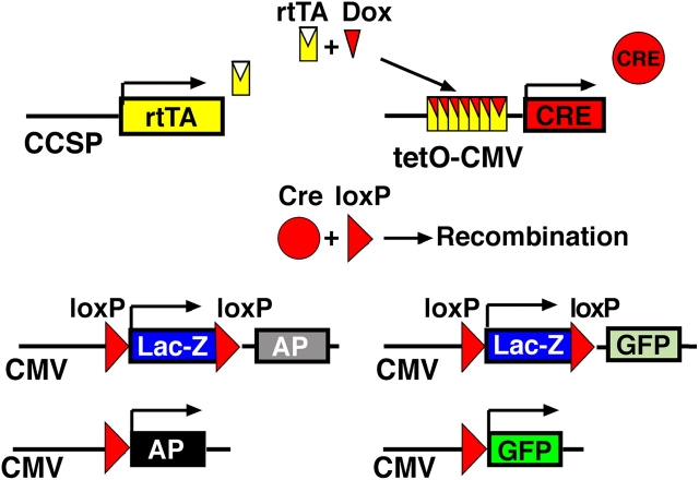
To identify relationships amongst tracheal and alveolar epithelial cells during lung development, we used conditional systems controlled by the rat CCSP and human SFTPC gene promoters to express Cre-recombinase in the developing mouse lung, thereby permanently labeling cells by expression of alkaline phosphatase or green fluorescent protein. When controlled by the rat CCSP promoter, continuous exposure of the fetus to doxycycline caused widespread recombination in conducting airway epithelial cells, including cells of the trachea, bronchi, and bronchioles before birth, and in both conducting and peripheral airways after birth. Neuroepithelial cells, identified by CGRP staining, were never labeled. Recombination and permanent labeling were observed in both ciliated and nonciliated respiratory epithelial cells, demonstrating their derivation from common progenitor cells during lung morphogenesis. Remarkable dorsal-ventral and cephalo-caudal labeling patterns, established before birth, were identified by recombination controlled by the rat CCSP gene promoter. In the trachea, subsets of epithelial cells labeled by the CCSP promoter were organized horizontally along the dorsal-ventral axis of the trachea, where selective labeling of cells juxtaposed to tracheal and bronchial cartilage was observed. In sharp contrast, recombination controlled by the human SFTPC gene promoter identified related cells that were organized in linear patterns along the cephalo-caudal axis of the conducting airways. Conditional expression of Cre-recombinase in the respiratory epithelium provides a useful model for the study of gene expression and function in the mouse respiratory tract and in the lung.


Figures

<b>Figure 1.</b>
Figure 1.
Permanent cell labeling by controlled expression of Cre-recombinase. The rtTA gene is driven by a cell-type specific promoter (rat CCSP) and followed by a SV40 polyadenylation site. In the presence of doxycycline (Dox), the transactivator (rtTA) recognizes its specific DNA target sequence (tetO). Expression of CRE causes recombination at loxP sites. In Z/AP and Z/EG reporter mice β-gal expression changes to alkaline phosphatase (AP) or enhanced green fluorescent protein (GFP). Visualization of AP or GFP expression indicates doxycycline-induced CCSP-dependent recombination in triple transgenic CCSPrtTA/tetOCre/ZAP and CCSPrtTA/tetOCre/ZEG mice.
<b>Figure 2.</b>
Figure 2.
Localization of rtTA mRNA in CCSPrtTA transgenic mice. Radioactive in situ hybridization was performed on paraffin sections of lung from CCSP-rtTA transgenic mice killed at E 14.5 (A), E 16.5 (B), E 18.5 (C), PN 5 (D), PN 7 (E), PN 10 (F), PN 14 (G), and PN 21 (H, I). The CCSP promoter drives rtTA expression in the bronchus and bronchioles as early as E 14.5 and in the alveolar region as early as E 18.5. In the trachea of adult mice, rtTA expression was detected primarily in epithelial cells on the ventral side of the trachea. V, ventral; D, dorsal.
<b>Figure 3.</b>
Figure 3.
Localization of endogenous CCSP mRNA in rat and mouse lung. Radioactive in situ hybridization was performed on paraffin sections of lung from adult rat (A, C) and mouse (B). Endogenous CCSP mRNA expression was detected in the bronchus, bronchioles, and a subset of alveolar type II cells in the rat (A). In the mouse, CCSP mRNA was detected in conducting airways but not in the alveoli (B). Hybridization with the sense RNA is shown on rat tissue (C). Size bars = 200 μm.
<b>Figure 4.</b>
Figure 4.
CCSPrtTA mediated recombination in adult and E 18.5 to PN 9 mouse lung. Lung sections of adult (A, B) and PN21 (C–H) double and triple transgenic mice were analyzed for AP expression. In the absence of doxycycline, rare recombination in the conducting airways was observed in lung sections of adult (arrows in A) and PN 21 (arrowhead in E, F) triple transgenic CCSPrtTA/tetOCRE/ZAP mice. No AP-positive cells were found in the lung periphery (A, F). No AP-positive cells were detected in double transgenic control lungs (C, D). After 3 wk of doxycycline treatment, recombination was found in ciliated cells, Clara cells (arrows in B), and a subset of type II cells (arrowheads in B) in triple transgenic adult CCSPrtTA/tetOCRE/ZAP mouse lung. After doxycycline exposure from E 18.5 to PN 9, most nonciliated and ciliated cells in the bronchioles were positive for AP staining in triple transgenic CCSPrtTA/tetOCRE/ZAP PN 18 lungs. Cil, ciliated cells; Cla, Clara cells. Size bars: A, B = 50 μm; C–H = 10 μm.
<b>Figure 5.</b>
Figure 5.
Recombination in embryonic lungs of CCSPrtTA/tetOCRE/ZAP mice after continuous exposure to doxycycline. Dams were treated with doxycycline from E 6.5 until killing. Mice were killed on E 14.5 (A), E 15.5 (B), E 16.5 (C), and at birth (D). Rare AP-positive cells were detected in conducting airways as early as E 14.5 (arrowhead), and in the lung parenchyma as early as E 16.5 (arrowhead). Numbers of labeled epithelial cells increased from E 14.5 until birth. Size bar = 50 μm.
<b>Figure 6.</b>
Figure 6.
Timing of recombination in the prenatal period. Dams were treated with doxycycline for 48-h periods. AP staining was assessed in triple transgenic CCSPrtTA/tetOCRE/ZAP lungs at E 18.5. When exposed to doxycycline from E 12.5–14.5, few cells were labeled in the bronchioles (A, D, G). After exposure from E 14.5–16.5, extensive labeling was observed in bronchioles and bronchiolar-alveolar portals, with a subset of cells labeled in peripheral saccules (B, E, H). After exposure from E 16.5–18.5, recombination was detected in most cells in the proximal and terminal bronchioles, but was rarely detected in peripheral lung saccules (C, F, I). Labeling was not detected in thymus or thyroid. Size bar = 100 μm.
<b>Figure 7.</b>
Figure 7.
Pattern of recombination from the trachea to the alveolar epithelium. Dams were treated with doxycycline from E 6.5 to PN 7. Tracheas of triple transgenic CCSPrtTA/tetOCRE/ZEG (A, C, E, G, I, K) and SPCrtTA/tetOCRE/ZEG (B, D, F, H, J, L) mice were visualized using an inverted microscope with fluorescence optics. Whole mount of the proximal trachea is shown in A and B. The main stem bronchi are shown in C and D. Inserts in A and C demonstrate the ventral (A) and dorsal (C) aspects of the trachea at 4× higher magnification. In CCSPrtTA/tetOCRE/ZEG tracheas, labeled cells were present in a dorsal–ventral pattern with increased density of labeled cells overlaying the cartilage rings. In the SPCrtTA/tetOCRE/ZEG tracheas, labeled cells formed longitudinal stripes with increasing numbers of labeled cells observed from the proximal to distal region. In the mainstem bronchi (E, G), labeled cells where found in a random pattern in the lungs of CCSPrtTA/tetOCRE/ZEG mice. Longitudinal stripes were observed in the bronchi of SPCrtTA/tetOCRE/ZEG mice (F, H). Recombination was frequent in epithelial cells of the bronchioles, as well as in the alveoli of both transgenic lines (I–L). Size bars: A, B = 400 μm; E, F = 500 μm; G, H, I, J = 250 μm; K, L = 400 μm. Note: autofluorescence can be seen in all tissue. V, ventral; D, dorsal.
<b>Figure 8.</b>
Figure 8.
Colocalization of GFP with β-tubulin and endogenous CCSP in the trachea. Dams were treated with doxycycline from E 6.5 to PN 7. Frozen sections of trachea (A, B) of triple transgenic CCSPrtTA/tetOCRE/ZEG mice were visualized with an upright microscope using fluorescence optics. Numerous GFP-positive cells were detected all along the ventral or cartilaginous side of the trachea (arrows in A) from the larynx to the mainstem bronchi (A, B). Tracheal glands lacked GFP staining (arrows in B). Frozen sections of CCSPrtTA/tetOCRE/ZEG trachea were stained for β-tubulin (C, D; red) or CCSP (E, F; red) and visualized for dual fluorescence with GFP (green). Ciliated cells on the dorsal side of the trachea were predominantly GFP-negative (C, arrows). The majority of ciliated cells (β-tubulin) on the ventral side of the trachea were labeled with GFP (D, arrow), although some ciliated cells were GFP-negative (C, D; arrowhead). CCSP did not colocalize with GFP anywhere in the trachea (E, F; arrowhead). Note: The yellow color on F results from close proximity of the red CCSP signal and the green GFP signal, but does not reflect colocalization. Arrows in all panels show GFP-negative cells that express differentiation markers; arrowheads demonstrate colocalization. Size bars: A, B = 500 μm; C, E = 200 μm; D, F = 50 μm.
<b>Figure 9.</b>
Figure 9.
Colocalization of GFP with epithelial cell markers in the peripheral lung. Dams were treated with doxycycline from E 6.5 to PN 7. Most bronchiolar cells (A) and some alveolar type II cells (B) expressed GFP. At PN 7 frozen sections of CCSPrtTA/tetOCRE/ZEG lungs were stained for β-tubulin, CCSP, pro–SP-C, or CGRP (red) and visualized for dual fluorescence with GFP (green). Some ciliated (β-tubulin–positive) cells expressed GFP (arrowhead in C). In the bronchioles, some Clara cells (CCSP-positive) expressed GFP (arrowhead in D), although not all nonciliated, GFP-expressing cells expressed CCSP (arrows in D). A subset of alveolar type II cells (pro–SP-C) expressed GFP (arrowhead in E). GFP was not detected in squamous type I epithelial cells (E). GFP expression (green) was not colocalized with CGRP (arrows in F). Note: arrows in all panels show GFP-negative cells that do express differentiation markers; arrowheads show colocalization. Size bars: A = 500 μm; B = 125 μm; C–F (left) = 50 μm; C–F (right) = 10 μm.

References

    1. Ten Have-Opbroek AA. Lung development in the mouse embryo. Exp Lung Res 1991;17:111–130. - PubMed
    1. Shannon JM. Induction of alveolar type II cell differentiation in fetal tracheal epithelium by grafted distal lung mesenchyme. Dev Biol 1994;166:600–614. - PubMed
    1. Cardoso WV. Molecular regulation of lung development. Annu Rev Physiol 2001;63:471–494. - PubMed
    1. Jeffery PK. Morphologic features of airway surface epithelial cells and glands. Am Rev Respir Dis 1983;128:S14–S20. - PubMed
    1. Mucenski ML, Wert SE, Nation JM, Loudy DE, Huelsken J, Birchmeier W, Morrisey EE, Whitsett JA. beta-Catenin is required for specification of proximal/distal cell fate during lung morphogenesis. J Biol Chem 2003;278:40231–40238. - PubMed

Publication types

MeSH terms