. 2005 Sep;139(1):186-91.
doi: 10.1104/pp.105.063529. Epub 2005 Jul 29.

Overexpression of TAPETUM DETERMINANT1 alters the cell fates in the Arabidopsis carpel and tapetum via genetic interaction with excess microsporocytes1/extra sporogenous cells

Affiliations

Overexpression of TAPETUM DETERMINANT1 alters the cell fates in the Arabidopsis carpel and tapetum via genetic interaction with excess microsporocytes1/extra sporogenous cells

Shu-Lan Yang et al. Plant Physiol. 2005 Sep.

Abstract

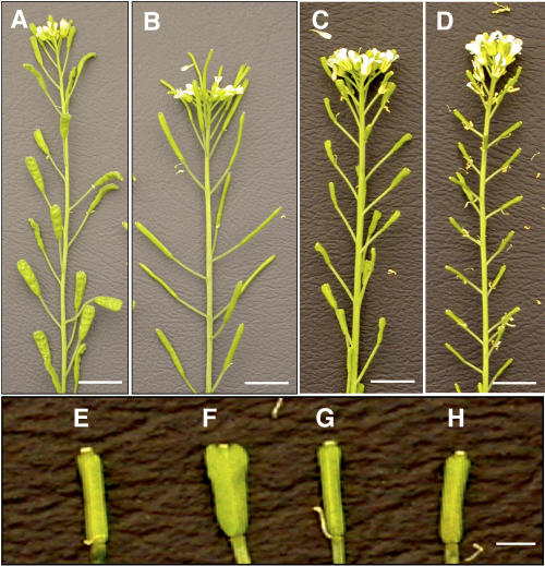
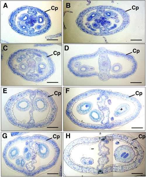
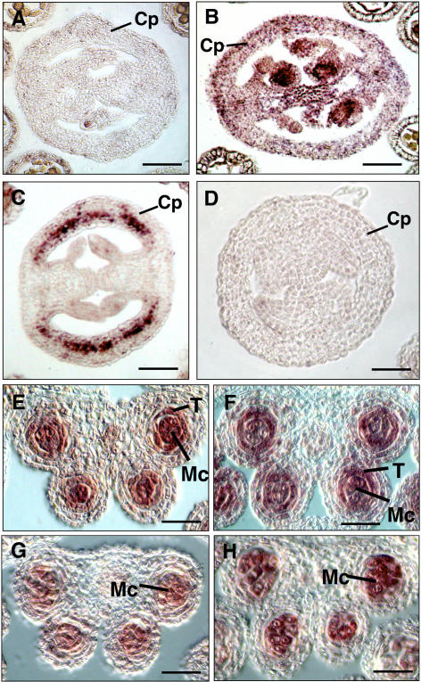
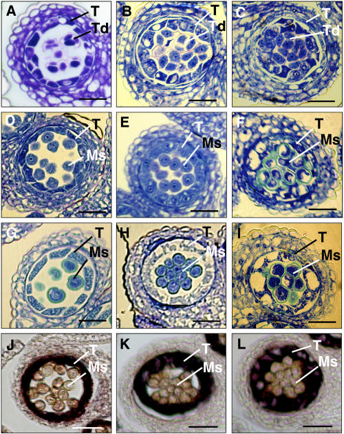
Previously, we reported that the TAPETUM DETERMINANT1 (TPD1) gene is required for specialization of tapetal cells in the Arabidopsis (Arabidopsis thaliana) anther. The tpd1 mutant is phenotypically identical to the excess microsporocytes1 (ems1)/extra sporogenous cells (exs) mutant. The TPD1 and EMS1/EXS genes may function in the same developmental pathway in the Arabidopsis anther. Here, we further report that overexpression of TPD1 alters the cell fates in the Arabidopsis carpel and tapetum. When TPD1 was expressed ectopically in the wild-type Arabidopsis carpel, the number of cells in the carpel increased significantly, showing that the ectopic expression of TPD1 protein could activate the cell division in the carpel. Furthermore, the genetic analysis showed that the activation of cell division in the transgenic carpel by TPD1 was dependent on EMS1/EXS, as it did not happen in the ems1/exs mutant. This result further suggests that TPD1 regulates cell fates in coordination with EMS1/EXS. Moreover, overexpression of TPD1 in tapetal cells also delayed the degeneration of tapetum. The TPD1 may function not only in the specialization of tapetal cells but also in the maintenance of tapetal cell fate.


Figures

Figure 1.
Figure 1.
Ectopic expression of TPD1 altered the pattern of siliques. A, A transgenic plant with wider siliques caused by ectopic expression of 35S::TPD1 in wild type. B, A wild-type plant. C, A transgenic plant with wider sterile siliques caused by expression of 35S::TPD1 in the tpd1 mutant. D, A tpd1 mutant plant. E, A tpd1 mutant silique. F, A transgenic sterile silique with a wider patterning, showing that ectopic expression of 35S::TPD1 altered the patterning of silique in the tpd1 mutant. G, A sterile ems1/exs-2 silique. H, A transgenic sterile silique with normal patterning, showing that the overexpression of 35S::TPD1 in ems1/exs-2 mutant did not alter the patterning of the silique. Bar = 1 cm in A to D; bar = 1 mm in E to H.
Figure 2.
Figure 2.
Comparison of the wider transgenic siliques to the wild-type siliques. The transversal sections show that the wider transgenic siliques have more cells than the wild-type siliques. A, There are 142.2 ± 10.4 epidermal cells in a transversal section of a wild-type silique at anthesis stage. B, There are 169.2 ± 14.2 epidermal cells in a transversal section of a wider transgenic silique at anthesis stage. C, There are 160.9 ± 11.9 epidermal cells in a transversal section of a wild-type silique at a stage 2 d after anthesis. D, There are 205.5 ± 17.5 epidermal cells in a transversal section of a wider transgenic silique at a stage 2 d after anthesis. E, There are 163.8 ± 10.5 epidermal cells in a transversal section of a wild-type silique at a stage 5 d after anthesis. F, There are 236.8 ± 20.1 epidermal cells in a transversal section of a wider transgenic silique at a stage 5 d after anthesis. G, There are 164.3 ± 12.8 epidermal cells in a transversal section of a wild-type silique at a stage 10 d after anthesis. H, There are 249.9 ± 20.1 epidermal cells in a transversal section of a wider transgenic silique at a stage 10 d after anthesis. Cp, Carpel. Bars = 25 μm.
Figure 3.
Figure 3.
Expression patterns of TPD1 in the siliques and anthers of wild-type, tpd1, ems1/exs-2, and transgenic plants. A, A transversal section of wild-type silique probed with the TPD1 antisense RNA. B, A transversal section of wild-type silique probed with the EMS1/EXS antisense RNA. C, A transversal section of the transgenic silique probed with the TPD1 antisense RNA. D, A transversal section of wild-type silique probed with the TPD1 sense RNA as a negative control. E, A wild-type anther section probed with the TPD1 antisense RNA. F, A wild-type anther section probed with the EMS1/EXS antisense RNA. G, An ems1 anther section probed with the TPD1 antisense RNA. H, A tpd1 anther section probed with the EMS1/EXS antisense RNA. Cp, Carpel; T, tapetum; Mc, microsporocyte. Bars = 20 μm.
Figure 4.
Figure 4.
Overexpression of TPD1 delayed the degeneration of transgenic tapetal cells. A to C, Transversal sections of anthers at stage 7, showing that tetrads were formed in the wild-type (A), fertile transgenic (B), and sterile transgenic (C) anthers. D to F, The transversal sections of anthers at stage 9. D, The tapetal cells started to degenerate and microspores were separated in wild-type anther. E, The degeneration of tapetal cells was delayed and microspores were still separated in the fertile transgenic anther. F, The degeneration of tapetal cells was greatly delayed and microspores were not separated in the sterile transgenic anther. G to I, The transversal sections of anthers at stage 11. G, The tapetal cells were almost degenerated and the pollen grains were formed in wild-type anther. H, The tapetal cells were degenerated apparently more slowly in the fertile transgenic anther than in the wild-type ones. The pollen grains were formed but stacked together. I, The degeneration of tapetal cells was greatly delayed and pollen grains were not separated in the sterile transgenic anther. J to L, Overexpression of TPD1 enhanced the expression of a tapetum-specific gene, ATA7, in the transgenic tapetal cells at stage 9, showing that the ATA7 was expressed more strongly in sterile (L) and fertile (K) transgenic anthers than in wild-type anther (J). Ms, Microspores; Pg, pollen grains; T, tapetum; Td, tetrads. Bars = 20 μm.

References

    1. Balasubramanian S, Schneitz K (2000) NOZZLE regulates proximal-distal pattern formation, cell proliferation and early sporogenesis during ovule development in Arabidopsis thaliana. Development 127: 4227–4238 - PubMed
    1. Bossinger G, Smyth D (1996) Initiation patterns of flower and floral organ development in Arabidopsis thaliana. Development 122: 1093–1102 - PubMed
    1. Brand U, Fletcher JC, Hobe M, Meyerowitz EM, Simon R (2000) Dependence of stem cell fate in Arabidopsis on a feedback loop regulated by CLV3 activity. Science 289: 617–619 - PubMed
    1. Canales C, Bhatt AM, Scott R, Dickkinson H (2002) EXS, a putative LRR receptor kinase, regulates male germline cell number and tapetal identity and promotes seed development in Arabidopsis. Curr Biol 12: 1718–1727 - PubMed
    1. Clark SE, Jacobsen SE, Levin JZ, Meyerowitz EM (1996) The CLAVATA and SHOOT MERISTEMLESS loci competitively regulate meristem activity in Arabidopsis. Development 122: 1567–1575 - PubMed

Publication types

Substances

Associated data