. 2005 Aug;25(16):6889-98.
doi: 10.1128/MCB.25.16.6889-6898.2005.

Semaphorin 4D/plexin-B1 induces endothelial cell migration through the activation of PYK2, Src, and the phosphatidylinositol 3-kinase-Akt pathway

Affiliations

Semaphorin 4D/plexin-B1 induces endothelial cell migration through the activation of PYK2, Src, and the phosphatidylinositol 3-kinase-Akt pathway

John R Basile et al. Mol Cell Biol. 2005 Aug.

Abstract

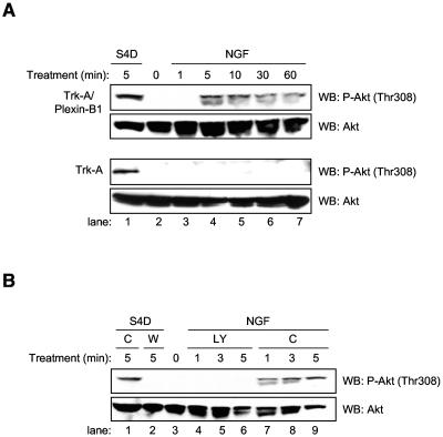
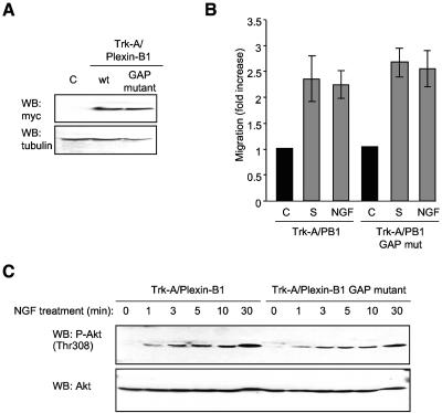
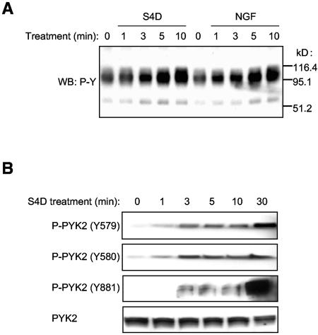
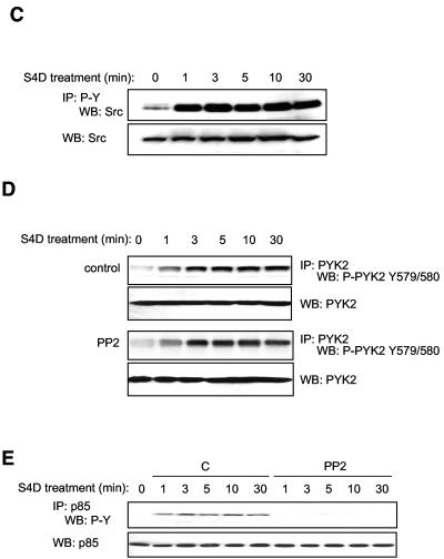
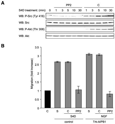
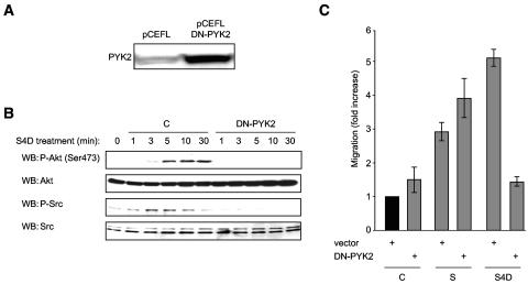
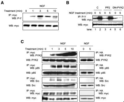
Semaphorins are cell surface and secreted proteins that provide axonal guidance in neuronal tissues and regulate cell motility in many cell types. They act by binding a family of transmembrane receptors known as plexins, which belong to the c-Met family of scatter factor receptors but lack an intrinsic tyrosine kinase domain. Interestingly, we have recently shown that Plexin-B1 is highly expressed in endothelial cells and that its activation by Semaphorin 4D elicits a potent proangiogenic response (J. R. Basile, A. Barac, T. Zhu, K. L. Guan, and J. S. Gutkind, Cancer Res. 64:5212-5224, 2004). In searches for the underlying molecular mechanism, we observed that Semaphorin 4D-stimulated endothelial cell migration requires the activation of the phosphatidylinositol 3-kinase (PI3K)-Akt pathway. Surprisingly, we found that Plexin-B1 stimulates PI3K-Akt through the activation of an intracellular tyrosine kinase cascade that involves the sequential activation of PYK2 and Src. This results in the tyrosine phosphorylation of Plexin-B1, the rapid recruitment of a multimeric signaling complex that includes PYK2, Src, and PI3K to Plexin-B1 and the activation of Akt. These findings suggest that Plexin-B1 may achieve its numerous physiological functions through the direct activation of intracellular tyrosine kinase cascades.


Figures

FIG. 1.
FIG. 1.
A role for the PI3K-Akt pathway in endothelial cell migration in response to Semaphorin 4D. (A) PI3K inhibition prevents Semaphorin 4D-mediated endothelial cell migration. Cells were preincubated with either DMSO vehicle control (−) or 50 μM of the PI3K inhibitor LY294002 (+) and used in a chemotaxis assay with purified (200 ng/ml) Semaphorin 4D as the chemoattractant (S4D). Media containing 10% fetal bovine serum (S) were used as positive controls for migration. The bars represent the fold increase of migration as determined by densitometry relative to that seen in negative control wells containing 0.1% BSA (C). (B) Endothelial cells were treated with 200 ng/ml Semaphorin 4D (S4D) for the indicated periods of time, lysed, and immunoprecipitated for the PI3K subunit p85 (IP: p85) and then immunoblotted for the presence of phosphorylated tyrosine residues (WB: P-Y). Phosphotyrosine was seen in p85 immunoprecipitates from Semaphorin 4D-treated cells starting at 1 min after treatment (upper panel). Total p85 was used as a loading control (WB: p85; lower panel). (C) Semaphorin 4D activates Akt in endothelial cells. Cells were treated with 200 ng/ml Semaphorin 4D (S4D) for the indicated periods of time, lysed, and analyzed for the presence of phosphorylated Akt. Phospho-Akt (P-Akt, Thr308) was seen in cells starting at 1 min after treatment (upper panel). Total Akt was used as a loading control (WB: Akt; lower panel). (D) Semaphorin 4D-mediated activation of Akt can be inhibited by LY294002 in endothelial cells. Cells preincubated with either vehicle control (C) or 50 μM LY294002 (LY) were treated with 200 ng/ml Semaphorin 4D (S4D) for the indicated periods of time, lysed, and analyzed for the presence of phosphorylated Akt. Phospho-Akt (P-Akt, Thr308) was seen in cells starting at 1 min after treatment in DMSO-pretreated cells but not in those treated with LY294002 (upper panel). Total Akt was used as a loading control (WB: Akt; lower panel).
FIG. 2.
FIG. 2.
NGF treatment of endothelial cells stably expressing Trk-A/Plexin-B1 chimeric receptor constructs induces Akt phosphorylation. (A) Cells stably expressing extracellular Trk-A or Trk-A fused to the intracellular portion of Plexin-B1 (Trk-A/Plexin-B1) were treated with 100 ng/ml NGF for the indicated periods of time, lysed, and analyzed for the presence of phosphorylated Akt (P-Akt, Thr308; upper panels). Cells treated with 200 ng/ml Semaphorin 4D for 5 min were used as a positive control (S4D; lane 1). Total Akt was used as a loading control (WB: Akt; lower panels). (B) NGF-induced phosphorylation of Akt in endothelial cells stably expressing Trk-A/Plexin-B1 chimeric receptor constructs can be inhibited by LY294002. Cells stably expressing Trk-A/Plexin-B1 chimeric receptors were preincubated with either 50 μM LY294002 (LY) or DMSO vehicle control (C) and treated with 100 ng/ml NGF for the indicated periods of time, lysed, and analyzed for the presence of phosphorylated Akt. Phospho-Akt (P-Akt, Thr308) was seen after treatment in control pretreated cells (lanes 7 to 9, upper panel) but not in those growing in LY294002 (lanes 4 to 6, upper panel). Cells treated with 200 ng/ml Semaphorin 4D and DMSO (C) or 100 nM Wortmannin (W) for 5 min were used as a positive control (lane 1) and negative control (lane 2), respectively. Total Akt was used as a loading control (WB: Akt, lower panel).
FIG. 3.
FIG. 3.
Endothelial cell migration and Akt phosphorylation and activation in response to Semaphorin 4D occur independently of residues involved in Plexin-B1 Ras GAP activity. (A) Transfected cells show immunoreactivity for the Ras GAP mutant form of the chimera Trk-A/Plexin-B1 (GAP mutant), at levels similar to that seen in cells expressing the wild-type intracellular portion of Plexin-B1 fused to Trk-A (wt, upper panel). Empty vector transfected cells were used as a negative control (C) and tubulin as the loading control (WB: tubulin, lower panel). (B) Endothelial cells expressing Trk-A/Plexin-B1 or Trk-A/Plexin-B1 Ras GAP mutant chimeras were used in a cell migration assay with 100 ng/ml NGF as the chemoattractant (NGF). Media containing 10% fetal bovine serum (S) was used as a positive control for migration. The bars represent the fold increase of migration as determined by densitometry relative to that seen in negative control wells containing 0.1% BSA (C). (C) Cells expressing Trk-A/Plexin-B1 or Trk-A/Plexin-B1 Ras GAP mutant chimeras were treated with 200 ng/ml Semaphorin 4D for the indicated periods of time, lysed, and analyzed for the presence of phosphorylated Akt. Phospho-Akt (WB: P-Akt [Thr308]) was seen in response to NGF treatment of Trk-A/Plexin-B1 expressing cells and in those expressing the Ras GAP mutant (upper panel). Total Akt was used as a loading control (WB: Akt, lower panel). mut, mutant.
FIG. 4.
FIG. 4.
Semaphorin 4D induces the tyrosine phosphorylation of PYK2 and Src in endothelial cells. (A) Treatment of endothelial cells with Semaphorin 4D and cells stably expressing Trk-A/Plexin-B1 chimeric receptor constructs with NGF induces changes in tyrosine phosphorylation. Endothelial cells and cells stably expressing Trk-A/Plexin-B1 chimeric receptors were treated with 200 ng/ml Semaphorin 4D (S4D) or 100 ng/ml NGF (NGF), respectively, for the indicated periods of time, lysed, and analyzed for the presence of tyrosine phosphoproteins (WB: P-Y). Changes in tyrosine phosphorylation of proteins of approximately 110 and 60 kDa are detected. (B) PYK2 is activated in endothelial cells in response to S4D. Endothelial cells were treated with 200 ng/ml Semaphorin 4D for the indicated periods of time, lysed, and immunoblotted with antibodies specific for different sites of tyrosine phosphorylation on PYK2. The levels of phosphorylation on Y579 and 580, residues that reflect kinase activity, and Y881, corresponding to the proposed Grb-2 binding site in the C-terminal domain, are observed starting at 1 min and 3 min after treatment with Semaphorin 4D, respectively. (C) Src is found in tyrosine-phosphorylated molecular complexes in endothelial cells in response to S4D. Cells were treated with 200 ng/ml Semaphorin 4D for the indicated periods of time, lysed, and immunoprecipitated for tyrosine-phosphorylated proteins. The detection of Src in the tyrosine immunoprecipitated fraction dramatically increased at 1 min after treatment (IP: P-Y and WB: Src; upper panel). Total Src was used as a loading control (WB: Src; lower panel). (D) Blockade of Src has no effect on Semaphorin 4D-induced PYK2 activation. Endothelial cells were preincubated with either DMSO vehicle control (C) or 10 μM PP2 (PP2) and treated with 200 ng/ml Semaphorin 4D for the indicated periods of time. Cells were lysed, immunoprecipitated for PYK2 (IP: PYK2), and immunoblotted for the presence of the phosphorylated tyrosine residues Y579 and Y580, located in the activation loop (WB: P-PYK2 Y579/580). Phosphorylated tyrosine residues were detected in the immunoprecipitated fraction starting at approximately 1 min after treatment in both control and PP2 treated populations. Total PYK2 was used as a loading control (WB: PYK2). (E) Blockade of Src prevents Semaphorin 4D-induced phosphorylation of p85 PI3K subunit. Endothelial cells were preincubated with either DMSO vehicle control (C) or 10 μM PP2 (PP2) and treated with 200 ng/ml Semaphorin 4D for the indicated periods of time. Cells were lysed, immunoprecipitated for p85, and immunoblotted for the presence of the phosphorylated tyrosine residues (IP: p85 and WB: P-Y). Phosphorylated tyrosine residues were detected in the immunoprecipitated fraction starting at approximately 1 min after treatment in the DMSO populations but not in cells incubated with PP2 (upper panel). Total p85 was used as a loading control (WB: p85; lower panel). MW, molecular mass.
FIG. 4.
FIG. 4.
Semaphorin 4D induces the tyrosine phosphorylation of PYK2 and Src in endothelial cells. (A) Treatment of endothelial cells with Semaphorin 4D and cells stably expressing Trk-A/Plexin-B1 chimeric receptor constructs with NGF induces changes in tyrosine phosphorylation. Endothelial cells and cells stably expressing Trk-A/Plexin-B1 chimeric receptors were treated with 200 ng/ml Semaphorin 4D (S4D) or 100 ng/ml NGF (NGF), respectively, for the indicated periods of time, lysed, and analyzed for the presence of tyrosine phosphoproteins (WB: P-Y). Changes in tyrosine phosphorylation of proteins of approximately 110 and 60 kDa are detected. (B) PYK2 is activated in endothelial cells in response to S4D. Endothelial cells were treated with 200 ng/ml Semaphorin 4D for the indicated periods of time, lysed, and immunoblotted with antibodies specific for different sites of tyrosine phosphorylation on PYK2. The levels of phosphorylation on Y579 and 580, residues that reflect kinase activity, and Y881, corresponding to the proposed Grb-2 binding site in the C-terminal domain, are observed starting at 1 min and 3 min after treatment with Semaphorin 4D, respectively. (C) Src is found in tyrosine-phosphorylated molecular complexes in endothelial cells in response to S4D. Cells were treated with 200 ng/ml Semaphorin 4D for the indicated periods of time, lysed, and immunoprecipitated for tyrosine-phosphorylated proteins. The detection of Src in the tyrosine immunoprecipitated fraction dramatically increased at 1 min after treatment (IP: P-Y and WB: Src; upper panel). Total Src was used as a loading control (WB: Src; lower panel). (D) Blockade of Src has no effect on Semaphorin 4D-induced PYK2 activation. Endothelial cells were preincubated with either DMSO vehicle control (C) or 10 μM PP2 (PP2) and treated with 200 ng/ml Semaphorin 4D for the indicated periods of time. Cells were lysed, immunoprecipitated for PYK2 (IP: PYK2), and immunoblotted for the presence of the phosphorylated tyrosine residues Y579 and Y580, located in the activation loop (WB: P-PYK2 Y579/580). Phosphorylated tyrosine residues were detected in the immunoprecipitated fraction starting at approximately 1 min after treatment in both control and PP2 treated populations. Total PYK2 was used as a loading control (WB: PYK2). (E) Blockade of Src prevents Semaphorin 4D-induced phosphorylation of p85 PI3K subunit. Endothelial cells were preincubated with either DMSO vehicle control (C) or 10 μM PP2 (PP2) and treated with 200 ng/ml Semaphorin 4D for the indicated periods of time. Cells were lysed, immunoprecipitated for p85, and immunoblotted for the presence of the phosphorylated tyrosine residues (IP: p85 and WB: P-Y). Phosphorylated tyrosine residues were detected in the immunoprecipitated fraction starting at approximately 1 min after treatment in the DMSO populations but not in cells incubated with PP2 (upper panel). Total p85 was used as a loading control (WB: p85; lower panel). MW, molecular mass.
FIG. 5.
FIG. 5.
Blockade of Src prevents Semaphorin 4D-induced Akt activation and cell migration. (A) PP2 blocks Src and Akt phosphorylation in Semaphorin 4D-treated endothelial cells. Endothelial cells were preincubated with either 10 μM PP2 (PP2) or DMSO (C) and treated with 200 ng/ml Semaphorin 4D for the indicated periods of time, lysed, and analyzed for the presence of phosphorylated Akt and Src. Phospho-Src (WB: P-Src) was seen after 1 min of treatment in cells preincubated with DMSO but not in cells grown in PP2 (upper panels). Total Src was used as a loading control (WB: Src). Phosphorylation of Akt is also seen in DMSO-treated control cells but not in PP2 treated populations [WB: P-Akt (Thr 308), lower panels]. Total Akt is used as a loading control (WB: Akt). (B) PP2 blocks Semaphorin 4D/Plexin-B1-mediated chemotaxis. Endothelial cells (control) or cells stably expressing Trk-A/Plexin-B1 chimeric receptors (Trk-A/PB1) were preincubated with either DMSO vehicle control (C) or 10 μM of the Src inhibitor PP2 (PP2) and then used in a cell migration assay with Semaphorin 4D (S4D) or 100 ng/ml NGF (NGF) as the chemoattractants. Media containing 10% fetal bovine serum (S) were used as positive controls for migration. The bars represent the fold increase of migration as determined by densitometry relative to that seen in negative control wells containing 0.1% BSA (C).
FIG. 6.
FIG. 6.
A dominant-negative form of PYK2 blocks Semaphorin 4D/Plexin-B1-mediated Akt and Src phosphorylation and chemotaxis. (A) Transfected cells show immunoreactivity for PYK2, indicating overexpression of the kinase-dead, dominant-negative version of PYK2 (DN-PYK2), compared to empty vector-transfected controls (pCEFL). (B) Cells transfected with empty vector control DNA (C) or kinase-dead dominant-negative PYK2 (DN-PYK2) were treated with 200 ng/ml Semaphorin 4D for the indicated periods of time, lysed, and analyzed for the presence of phosphorylated Akt and Src. Phospho-Akt [WB: P-Akt (Ser 473); upper panels] and phospho-Src (WB: P-Src; lower panels) were seen in response to Semaphorin 4D treatment of control cells but not in those expressing DN-PYK2. Total Akt and Src were used as loading controls. (C) Endothelial cells transfected with empty vector control DNA (vector) or kinase-dead dominant-negative PYK2 (DN-PYK2) were used in a cell migration assay with 200 ng/ml Semaphorin 4D as the chemoattractant (S4D). Media containing 10% fetal bovine serum (S) were used as positive controls for migration. The bars represent the fold increase of migration as determined by densitometry relative to that seen in negative control wells containing 0.1% BSA (C).
FIG. 7.
FIG. 7.
Plexin-B1 activation promotes the assembly of multimeric signaling complexes. (A) Plexin-B1 is tyrosine phosphorylated upon activation. Lysates of endothelial cells stably expressing myc-tagged Trk-A/Plexin-B1 chimeric receptors treated with 100 ng/ml NGF for the indicated periods of time were immunoprecipitated for the receptor (IP: myc) and immunoblotted for the presence of phosphorylated tyrosine residues (WB: P-Y). A phosphotyrosine band of the appropriate size begins to appear in the immunoprecipitated fraction at about 1 min (upper panel). Total receptor (WB: myc) was used as a loading control (lower panel). (B) 293T cells were cotransfected with myc-tagged Trk-A/Plexin-B1 and either DN-PYK2 (DN-PYK2), control DNA (C), or control DNA preceded by incubation with the Src inhibitor PP2 (PP2), followed by treatment with 100 ng/ml NGF. An immunoblot for myc was performed on phosphotyrosine-immunoprecipitated fractions (IP: P-Y and WB: myc). An increase in receptor phosphorylation is seen in response to NGF in control cells (lane 2) and cells pretreated with PP2 (lane 4) but not in cells expressing DN-PYK2 (lane 6). (C) PYK2, p85, and Src bind Plexin-B1 upon receptor activation. Lysates of endothelial cells stably expressing myc-tagged Trk-A/Plexin-B1 chimeric receptors treated with 100 ng/ml NGF for the indicated periods of time were immunoprecipitated for the receptor (IP: myc) and immunoblotted for the presence of PYK2, p85, and Src. Each of these proteins is seen associating with the immunoprecipitated receptor complex at 1 min following treatment. Total PYK2, p85, Src, and myc immunoblots were used as loading controls. As a negative control, a hemagglutinin immunoprecipitation was performed (IP: HA; right panels) which showed no proteins after 0 or 5 min treatment with NGF when probed for PYK2, p85, Src, and myc. Total PYK2, p85, Src, and myc were used as loading controls (WB lanes; lower panels).

References

    1. Alessi, D. R., M. Andjelkovic, B. Caudwell, P. Cron, N. Morrice, P. Cohen, and B. A. Hemmings. 1996. Mechanism of activation of protein kinase B by insulin and IGF-1. EMBO J. 15:6541-6551. - PMC - PubMed
    1. Anonymous. 1999. Unified nomenclature for the semaphorins/collapsins. Semaphorin Nomenclature Committee. Cell 97:551-552. - PubMed
    1. Aurandt, J., H. G. Vikis, J. S. Gutkind, N. Ahn, and K. L. Guan. 2002. The semaphorin receptor plexin-B1 signals through a direct interaction with the Rho-specific nucleotide exchange factor, LARG. Proc. Natl. Acad. Sci. USA 99:12085-12090. - PMC - PubMed
    1. Avraham, H., S. Y. Park, K. Schinkmann, and S. Avraham. 2000. RAFTK/Pyk2-mediated cellular signalling. Cell Signal. 12:123-133. - PubMed
    1. Basile, J. R., A. Barac, T. Zhu, K. L. Guan, and J. S. Gutkind. 2004. Class IV semaphorins promote angiogenesis by stimulating Rho-initiated pathways through plexin-B. Cancer Res. 64:5212-5224. - PubMed

MeSH terms