. 2005 Aug;25(16):7181-92.
doi: 10.1128/MCB.25.16.7181-7192.2005.

Signal regulatory protein alpha ligation induces macrophage nitric oxide production through JAK/STAT- and phosphatidylinositol 3-kinase/Rac1/NAPDH oxidase/H2O2-dependent pathways

Affiliations

Signal regulatory protein alpha ligation induces macrophage nitric oxide production through JAK/STAT- and phosphatidylinositol 3-kinase/Rac1/NAPDH oxidase/H2O2-dependent pathways

Jacqueline Alblas et al. Mol Cell Biol. 2005 Aug.

Abstract

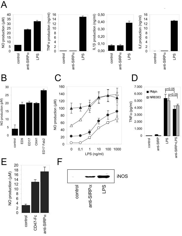
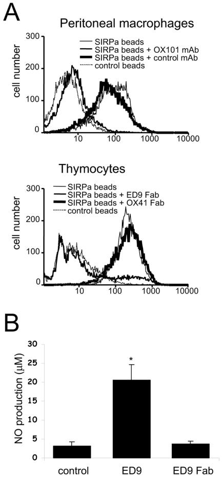
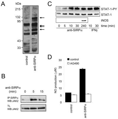
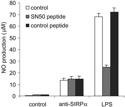
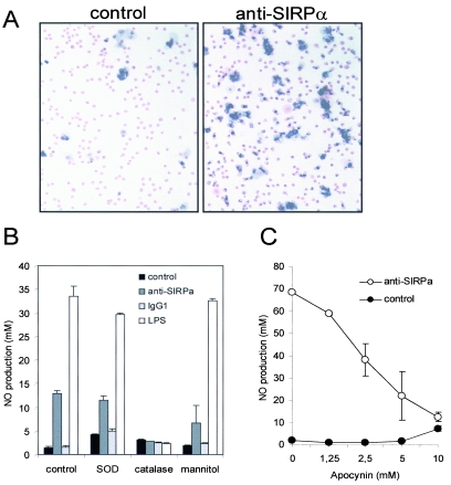
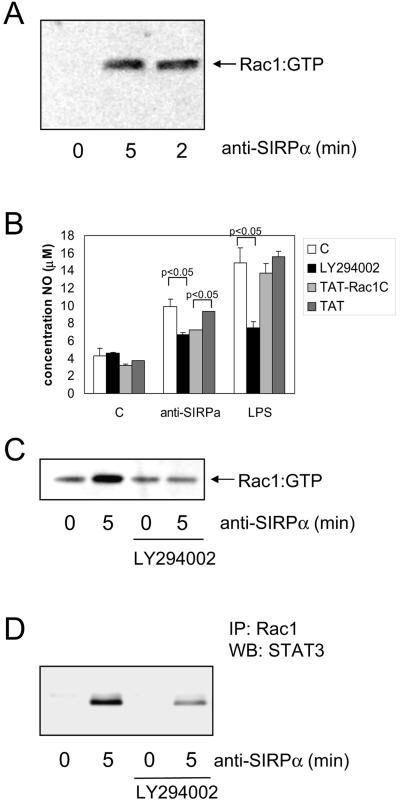
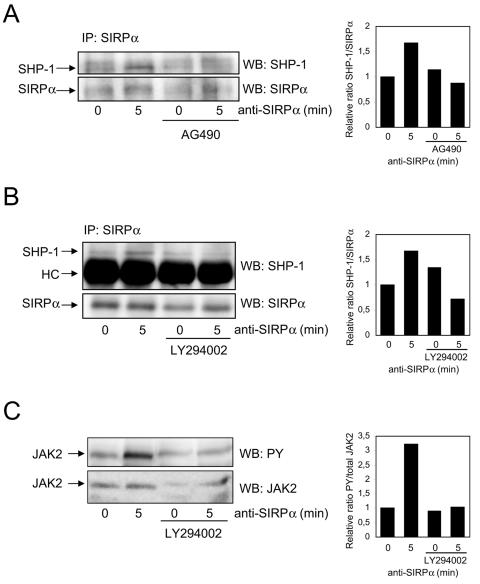
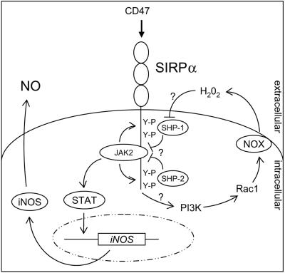
Signal regulatory protein alpha (SIRPalpha) is a glycoprotein receptor that recruits and signals via the tyrosine phosphatases SHP-1 and SHP-2. In macrophages SIRPalpha can negatively regulate the phagocytosis of host cells and the production of tumor necrosis factor alpha. Here we provide evidence that SIRPalpha can also stimulate macrophage activities, in particular the production of nitric oxide (NO) and reactive oxygen species. Ligation of SIRPalpha by antibodies or soluble CD47 triggers inducible nitric oxide synthase expression and production of NO. This was not caused by blocking negative-regulatory SIRPalpha-CD47 interactions. SIRPalpha-induced NO production was prevented by inhibition of the tyrosine kinase JAK2. JAK2 was found to associate with SIRPalpha in macrophages, particularly after SIRPalpha ligation, and SIRPalpha stimulation resulted in JAK2 and STAT1 tyrosine phosphorylation. Furthermore, SIRPalpha-induced NO production required the generation of hydrogen peroxide (H(2)O(2)) by a NADPH oxidase (NOX) and the phosphatidylinositol 3-kinase (PI3-K)-dependent activation of Rac1, an intrinsic NOX component. Finally, SIRPalpha ligation promoted SHP-1 and SHP-2 recruitment, which was both JAK2 and PI3-K dependent. These findings demonstrate that SIRPalpha ligation induces macrophage NO production through the cooperative action of JAK/STAT and PI3-K/Rac1/NOX/H(2)O(2) signaling pathways. Therefore, we propose that SIRPalpha is able to function as an activating receptor.


Figures

FIG. 1.
FIG. 1.
Ligation of SIRPα induces macrophage NO production. (A) Rat NR8383 macrophages were cultured for 18 h. in the presence of control IgG1 (MAb BF5, 20 μg/ml), anti-SIRPα MAb ED9 (20 μg/ml), or LPS (100 ng/ml), and amounts of secreted NO, TNF-α, IL-1β, or IL-6 were determined. (B) NO production in NR8383 macrophages was measured after addition of different intact MAb or corresponding F(ab)2 fragments (all at 20 μg/ml; 18 h of incubation). (C) Effects of control IgG1 (20 μg/ml, ○), anti-SIRPα MAb ED9 (20 μg/ml, •), IFN-γ (20 U/ml, ▵), or ED9 plus IFN-γ (▴) either alone, or in combination with various LPS concentrations, on NO production in rat peritoneal macrophages. (D) TNF-α secretion by peritoneal macrophages (PMph) or NR8383 cells cultured in the presence or absence of ED9 (20 μg/ml) and/or LPS (100 ng/ml) for 18 h (P values were obtained by Student's t test). (E) NO production in NR8383 macrophages triggered by murine CD47-Fc protein (25 μg/ml; 18 h of incubation) or anti-SIRPα MAb ED9 (10 μg/ml). (F) Anti-SIRPα MAb ED9 (10 μg/ml; 18 h of incubation) induces iNOS protein expression in NR8383 macrophages as shown by Western blotting. Experiments were performed at least in triplicate, and results are shown as means ± SD.
FIG. 2.
FIG. 2.
SIRPα-induced NO production by macrophages does not involve blocking of SIRPα-CD47 interactions. (A) Flow cytometric analysis of the binding of beads coated with rat SIRPα-CD4d3 + 4 chimeric protein (or control CD4d3 + 4 beads) to rat peritoneal macrophages or rat thymocytes. Where indicated, cells were preincubated with anti-CD47 MAb OX101 or control IgG1 (MAb OX45) or with anti-SIRPα ED9 Fab fragments or OX41 Fab fragments. (B) NO production by NR8383 cells stimulated with intact ED9 MAb (10 μg/ml) or ED9 Fab fragments (20 μg/ml). Data are expressed as the means ± SD from three independent experiments (*, P < 0.01 by Student's t test).
FIG. 3.
FIG. 3.
Role of JAK2/STAT signaling in SIRPα-induced NO production in NR8383 macrophages. (A) Tyrosine phosphorylation in macrophages stimulated for 5 min with control IgG1 (MAb BF5, 20 μg/ml) or anti-SIRPα MAb ED9 (20 μg/ml). Total lysates of NR8383 cells subjected to Western blotting and staining with anti-PY MAb 4G10. Note that SIRPα ligation promotes tyrosine phosphorylation of several cellular proteins (arrows). (B) Immunoprecipitates (IP) of SIRPα in NR8383 cells after stimulation by anti-SIRPα MAb ED9 (10 μg/ml) or total lysates (TL) were Western blotted (WB) and probed with anti-JAK2. Note that JAK2 associates with SIRPα in particular after SIRPα ligation by anti-SIRPα MAb ED9 (10 μg/ml). (C) Anti-SIRPα MAb ED9 (10 μg/ml)- or IFN-γ (20 U/ml)-stimulated NR8383 cells were lysed directly in SDS sample buffer. The panels show the same Western blot probed sequentially with anti-STAT1-PY, total anti-STAT1, and anti-iNOS antibodies. (D) Anti-SIRPα (MAb ED9, 20 μg/ml, 18 h of incubation)-induced NO production with or without 30 min of preincubation with AG490 (10 μM). Results from representative experiment that was performed in triplicate are shown (results are expressed as means ± SD).
FIG. 4.
FIG. 4.
Effect of NF-κB inhibitor on SIRPα-induced NO production. Anti-SIRPα (MAb ED9, 20 μg/ml)- or LPS (100 ng/ml)-induced NO production (18 h incubation) in the absence or presence of cell permeative, NF-κB-inhibitory SN50 peptide or inactive control peptide (50 μg/ml, added 30 min before ED9/LPS) is shown. The experiment was performed in triplicate, and results are shown as means ± SD.
FIG. 5.
FIG. 5.
SIRPα-induced triggering of the oxidative burst and the role of H2O2 in SIRPα-mediated NO production. (A) Superoxide production in rat peritoneal macrophages analyzed by NBT assay in the presence or absence of control IgG1 (MAb BF5, 20 μg/ml) or anti-SIRPα MAb (ED9, 20 μg/ml) for 90 min. (B) Effect of the ROS scavengers SOD (104 U/ml), catalase (104 U/ml), and mannitol (10 mM) on SIRPα-induced (ED9, 20 μg/ml, 18 h) NO production in NR8383 cells. (C) SIRPα MAb ED9-induced production of NO in NR8383 cells in the presence of the NADPH oxidase inhibitor apocynin. All experiments were performed in triplicate, and results are shown as means ± SD.
FIG. 6.
FIG. 6.
Role of Rac1 and PI3-K in SIRPα-induced NO production in macrophages. (A) Rac1 activation was determined (see Materials and Methods for details) in NR8383 cells following SIRPα ligation using MAb ED9 (10 μg/ml). (B) NR8383 cells in the presence of LY294002 (40 μM; 30-min preincubation), cell permeable TAT-Rac1 C-terminal peptides (200 μM; 5-min preincubation), or control TAT peptide (200 μM) were subsequently stimulated by anti-SIRPα (10 μg/ml) or LPS (100 ng/ml). After 20 h, NO production in the supernatants was measured. Shown are results from a representative experiment performed in triplicate (means ± SD). The differences between control and LY294002 in the anti-SIRPα- and LPS-treated samples and between Tat and Tat-Rac1 in the anti-SIRPα-treated samples are statistically significant (P < 0.05 by Student's t test). (C and D) NR8383 cells were pretreated for 30 min with LY294002 (40 μM) and then stimulated with anti-SIRPα (10 μg/ml) for 5 min and lysed. From these lysates, active Rac1 was pulled down as described in Materials and Methods (C), and subsequently, total Rac1 was immunoprecipitated (IP) and the associated STAT3 shown on a Western blot (WB) (D). Shown are representative results from three experiments.
FIG. 7.
FIG. 7.
Ligation of SIRPα promotes the JAK- and PI3-K-dependent recruitment of SHP-1 in macrophages. NR8383 cells were preincubated with AG490 (10 μM; 30 min) or LY294002 (40 μM; 30 min), control cells were incubated with vehicle, and then cells were stimulated with anti-SIRPα (10 μg/ml ED9) for 5 min, lysed, and immunoprecipitated (IP) with anti-SIRPα Abs (rabbit anti-SIRPα from PharMingen in panels A and B) or directly lysed in reducing SDS sample buffer (C). Western blots (WB) of these samples were stained with the indicated antibodies recognizing SHP-1, SHP-2, SIRPα (Santa Cruz Ab), PY, or JAK2 as described in Materials and Methods. HC, heavy chain.
FIG. 8.
FIG. 8.
Model for the signaling events that mediate SIRPα-induced NO production in macrophages. SIRPα oligomerization by ligand triggers cross-phosphorylation of SIRPα-associated JAK2 and promotes SIRPα tyrosine phosphorylation, which then allows STAT docking and tyrosine phosphorylation. Activated STAT dimers induce iNOS gene expression and subsequent NO production. Simultaneously, SIRPα ligation triggers Rac1 activation, in a PI3-K-dependent fashion, followed by NOX assembly and oxidative burst generation. The resultant H2O2 inhibits the catalytic activity of SIRPα-associated SHP-1 and SHP-2, thereby preventing JAK2 and SIRPα dephosphorylation resulting in sustained JAK-STAT signaling and enhanced SHP-1 and SHP-2 retention by SIRPα.

References

    1. Adams, S., L. J. van der Laan, E. Vernon-Wilson, C. Renardel de Lavalette, E. A. Dopp, C. D. Dijkstra, D. L. Simmons, and T. K. van den Berg. 1998. Signal-regulatory protein is selectively expressed by myeloid and neuronal cells. J. Immunol. 161:1853-1859. - PubMed
    1. Bright, J. J., C. Natarajan, S. Sriram, and G. Muthian. 2004. Signaling through JAK2-STAT5 pathway is essential for IL-3-induced activation of microglia. Glia 45:188-196. - PubMed
    1. Brooke, G., J. D. Holbrook, M. H. Brown, and A. N. Barclay. 2004. Human lymphocytes interact directly with CD47 through a novel member of the signal regulatory protein (SIRP) family. J. Immunol. 173:2562-2570. - PubMed
    1. Brown, M. H., K. Boles, P. A. van der Merwe, V. Kumar, P. A. Mathew, and A. N. Barclay. 1998. 2B4, the natural killer and T cell immunoglobulin superfamily surface protein, is a ligand for CD48. J. Exp. Med. 188:2083-2090. - PMC - PubMed
    1. de Vries, H. E., J. J. Hendriks, H. Honing, C. R. De Lavalette, S. M. van der Pol, E. Hooijberg, C. D. Dijkstra, and T. K. van den Berg. 2002. Signal-regulatory protein alpha-CD47 interactions are required for the transmigration of monocytes across cerebral endothelium. J. Immunol. 168:5832-5839. - PubMed

MeSH terms

Substances