. 2005 Aug;25(16):7203-15.
doi: 10.1128/MCB.25.16.7203-7215.2005.

Surveillance of different recombination defects in mouse spermatocytes yields distinct responses despite elimination at an identical developmental stage

Affiliations

Surveillance of different recombination defects in mouse spermatocytes yields distinct responses despite elimination at an identical developmental stage

Marco Barchi et al. Mol Cell Biol. 2005 Aug.

Abstract

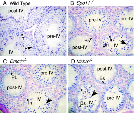
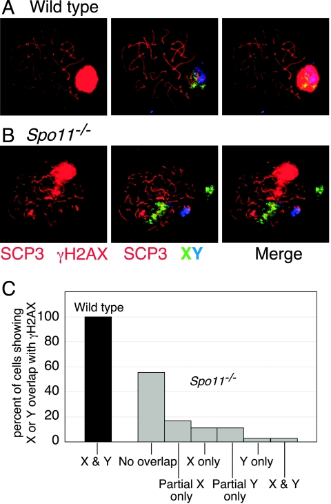
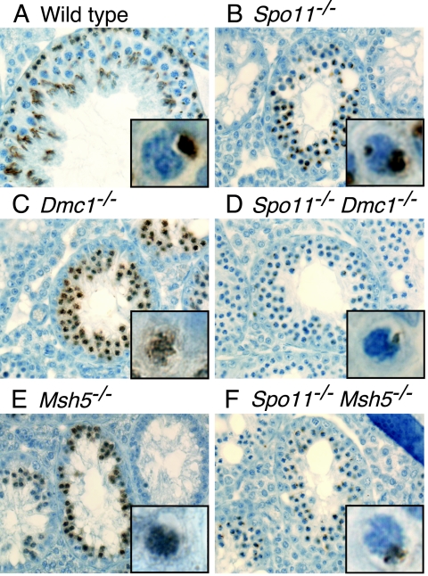
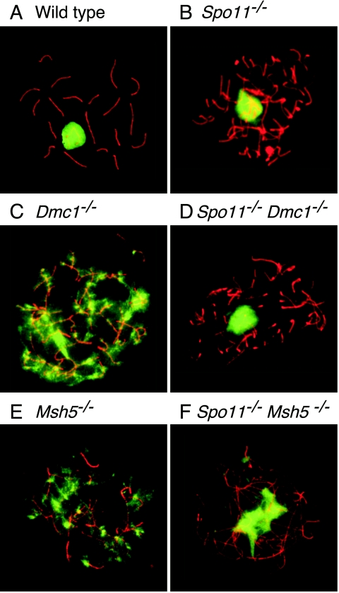
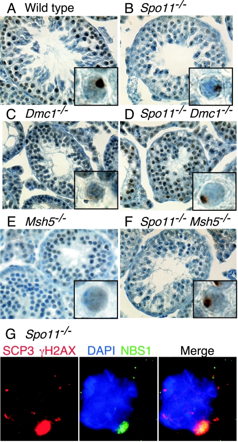
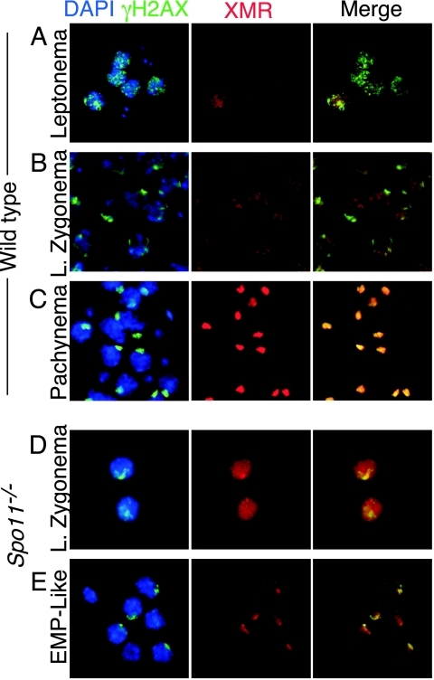
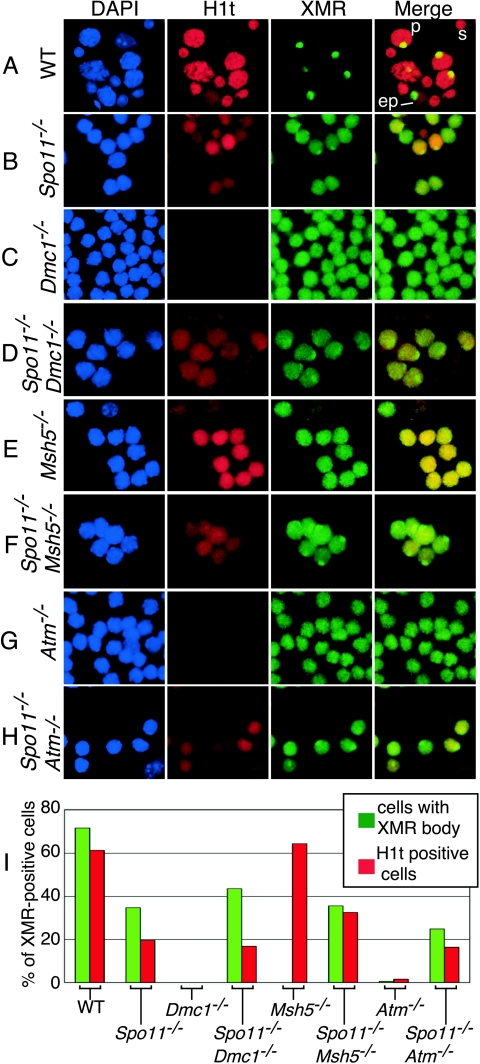
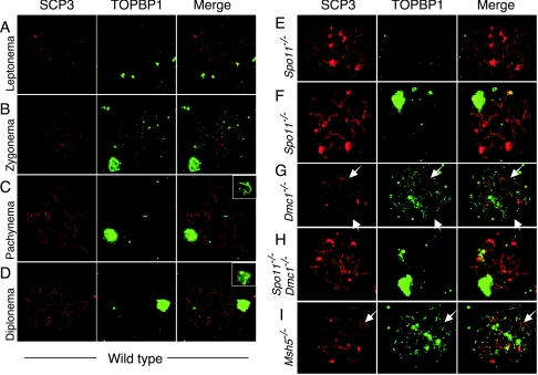
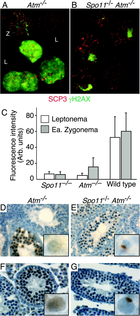
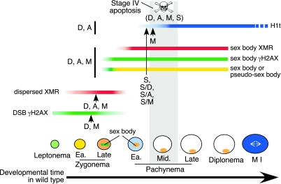
Fundamentally different recombination defects cause apoptosis of mouse spermatocytes at the same stage in development, stage IV of the seminiferous epithelium cycle, equivalent to mid-pachynema in normal males. To understand the cellular response(s) that triggers apoptosis, we examined markers of spermatocyte development in mice with different recombination defects. In Spo11(-)(/)(-) mutants, which lack the double-strand breaks (DSBs) that initiate recombination, spermatocytes express markers of early to mid-pachynema, forming chromatin domains that contain sex body-associated proteins but that rarely encompass the sex chromosomes. Dmc1(-)(/)(-) spermatocytes, impaired in DSB repair, appear to arrest at or about late zygonema. Epistasis analysis reveals that this earlier arrest is a response to unrepaired DSBs, and cytological analysis implicates the BRCT-containing checkpoint protein TOPBP1. Atm(-)(/)(-) spermatocytes show similarities to Dmc1(-)(/)(-) spermatocytes, suggesting that ATM promotes meiotic DSB repair. Msh5(-)(/)(-) mutants display a set of characteristics distinct from these other mutants. Thus, despite equivalent stages of spermatocyte elimination, different recombination-defective mutants manifest distinct responses, providing insight into surveillance mechanisms in male meiosis.


Figures

FIG. 1.
FIG. 1.
Different recombination defects trigger similar timings of spermatocyte apoptosis. (A to D) Periodic acid-Schiff-stained testis sections from wild-type and the indicated mutant mice. Examples of tubules at epithelial stage IV and the stages before (pre-IV) and after (post-IV) stage IV are indicated. Apoptotic cells (arrows) were observed at epithelial stage IV in the mutants, when pachytene spermatocytes (P) are normally present. Earlier-stage spermatocytes present in the mutants are indicated (preleptotene, PL; leptotene, L), as are intermediate (In) and B (Bs) spermatogonia.
FIG. 2.
FIG. 2.
Spo11/ spermatocytes form pseudo-sex bodies. (A and B) Immunofluorescence and FISH staining of spermatocyte spreads. In wild-type spermatocytes, the X and Y chromosomes colocalize with a discrete domain of γH2AX staining (the sex body). In Spo11/ spermatocytes the sex chromosomes rarely localize to the domain of γH2AX staining, revealing that this is not a true sex body. (C) Quantification of the frequency of overlap of the sex chromosomes with the γH2AX domain in wild-type and Spo11/ spermatocytes.
FIG. 3.
FIG. 3.
Localized accumulation of γH2AX does not occur in recombination mutants that are defective in the repair of SPO11-generated DSBs. Testis sections were stained with anti-γH2AX (brown). Insets show higher-magnification views of individual spermatocytes. As in the wild type (A), the γH2AX signal occurred in discrete domains in Spo11/ spermatocytes that had progressed sufficiently far in meiotic prophase (B). However, a subset of Spo11/ spermatocytes contained two localized domains of γH2AX, indicating that sex body formation did not always appear normal in this mutant (inset in panel B). In Dmc1/ (C) and Msh5/ (E) spermatocytes, γH2AX was distributed across most of the chromatin. This γH2AX localization defect was suppressed by the introduction of a Spo11 mutation (D and F).
FIG. 4.
FIG. 4.
Mutants defective in the repair of SPO11-generated DSBs do not form sex body-like domains of γH2AX. Chromosome spreads were stained with anti-SCP3 (red) and anti-γH2AX (green). The γH2AX signal occurred in discrete domains in wild-type (A) and in Spo11/ (B) spermatocytes but was distributed in patches across most of the chromatin in Dmc1/ (C) and Msh5/ (E) spermatocytes. The defect in γH2AX localization in Dmc1/ and Msh5/ spermatocytes was suppressed by Spo11 mutation (panels D and F, respectively).
FIG. 5.
FIG. 5.
Localized accumulation of NBS1 does not occur in recombination mutants that are defective in the repair of SPO11-generated DSBs. (A to F) Testis sections were stained with anti-NBS1 (brown). As in the wild type (panel A), the NBS1 signal occurred in discrete domains in Spo11/ spermatocytes that had progressed sufficiently far in meiotic prophase (panel B). In Dmc1/ (panel C) and Msh5/ (panel E) spermatocytes, NBS1 did not localize to discrete domains, although the NBS1 localization defects in Dmc1/ and Msh5/ spermatocytes were suppressed by the introduction of a Spo11 mutation (panels D and F, respectively). (G) Immunofluorescence staining of a spermatocyte spread with anti-γH2AX and anti-SCP3 in red and anti-NBS1 in green. NBS1 is a component of the sex body (here, the pseudo-sex body), as indicated by its colocalization with γH2AX.
FIG. 6.
FIG. 6.
Staining patterns of γH2AX and XMR in wild-type and Spo11/ spermatocytes in squash preparations of testicular cells. Squash preparations of testicular cells were stained with DAPI (blue), anti-γH2AX (green), and anti-XMR (red). (A to C) Staining patterns in wild-type cells, as previously described (27, 47). At leptonema (panel A), γH2AX is present in focal regions across the chromatin, and XMR staining is very weak. By late (L.) zygonema, γH2AX localization to the sex body becomes apparent, while XMR staining is diffuse (B). By pachynema, both markers colocalize to the sex body (C). (D and E) Staining patterns in Spo11/ spermatocytes. At late zygonema, γH2AX is localized to a discrete domain, the pseudo-sex body (D), while XMR staining is diffuse. By the early to mid-pachynema (EMP)-like stage, both markers colocalize (E).
FIG. 7.
FIG. 7.
Different recombination mutants show distinct patterns of markers for meiotic progression. (A to H) Squash preparations of testicular cells from mice of the indicated genotypes were stained for histone H1t and XMR. Examples of early pachytene (ep) and mid- to late-pachytene (p) spermatocytes and of a spermatid (s) are shown for the wild type (WT) (A). Some Spo11/ spermatocytes (B) stain positive for H1t, although generally not as brightly as the wild type, and localize XMR to a discrete domain, although in fewer cells than in the wild type. In Dmc1/ spermatocytes (C), histone H1t deposition was not detected and XMR was diffuse on chromatin, never accumulating in discrete patches. Spo11/ Dmc1/ spermatocytes (D) gave H1t and XMR staining patterns similar to those of Spo11/ spermatocytes. In Msh5/ spermatocytes (E), histone Hlt straining was de-tected but XMR was only diffuse. Spo11/ Msh5/ spermatocytes (F) gave H1t and XMR staining patterns similar to those of Spo11/ spermatocytes. Atm/ spermatocytes behaved similarly to Dmc1/ spermatocytes, both for the single mutant (G) and when combined with the Spo11/ mutation (H). (I) XMR-positive cells were counted for those with XMR concentrated in a discrete domain and those that stained positive for histone H1t. The absence of a bar indicates that the frequency of cells in that class was zero (see text).
FIG. 8.
FIG. 8.
Association of TOPBP1 with meiotic chromosomes in Dmc1/ and Msh5/ spermatocytes but not in Spo11/ spermatocytes. Spermatocyte chromosome spreads were stained with anti-SCP3 (red) and anti-TOPBP1 (green). Merged images are also shown. (A to D) In wild-type cells, TOPBP1 forms foci along the axes of chromosomes in leptonema (A) and zygonema (B), and the foci disappear or are greatly reduced on autosomes as DSBs are repaired and chromosomes synapse (C) in a manner similar that previously observed with a different antibody (33). TOPBP1 protein persists on the unsynapsed axes and chromatin of the XY pair from pachynema into diplonema (B to D). Note that the XY pair in panel B is from an adjacent pachytene nucleus. (E and F) In Spo11/ spermatocytes, TOPBP1 staining was virtually absent from most of the chromatin, except for the pseudo-sex body when present (F). (G to I) In Dmc1/ spermatocytes, TOPBP1 accumulated to levels higher than those in the wild type and persisted on chromosome axes and surrounding chromatin without forming obvious pseudo-sex bodies (G). Spo11/ Dmc1/ double mutants were indistinguishable from Spo11/ single mutants (H). Msh5/ spermatocytes (I) accumulated TOPBP1 in a fashion similar to that of Dmc1/ spermatocytes. TOPBP1 foci were absent or reduced on many axes undergoing at least limited synaptonemal complex formation (arrows in panels G and I). Matched exposures are shown, except for the insets to panels C and D, which show lower exposures of the TOPBP1 staining in the sex bodies.
FIG. 9.
FIG. 9.
Atm/ spermatocytes show hallmarks of the presence of poorly repaired DSBs. (A and B) Collages of chromosome spreads from Atm/ and Spo11/ Atm/ testes, stained with anti-SCP3 (red) and anti-γH2AX (green). Atm/ leptotene cells (L) showed little or no γH2AX signal, comparable to Spo11/ cells. Zygotene cells (Z) had a γH2AX signal that was above that of Spo11/ cells although reduced compared to that of the wild type. Atm/ spermatocytes were also observed with abundant γH2AX signal dispersed across the chromatin (bright green staining). Spo11/ Atm/ double mutants were indistinguishable from Spo11/ single mutants. (C) Fluorescence intensity (in arbitrary [Arb.] units) of the γH2AX signal of Atm/ compared to those of Spo11/ and wild-type leptotene andearly (Ea.) zygotene spermatocytes. The γH2AX (bright) Atm/ cells were excluded from this quantification. (D to G) Testis sections of Atm/ and Spo11/ Atm/ mice stained for γH2AX (D and E) or NBS1 (F and G). Insets show higher-magnification views of individual spermatocytes. The defect in sex body formation in Atm/ cells is apparent in these sections; the Spo11/ mutation rescues this defect to allow the localization of both γH2AX and NBS1 to pseudo-sex bodies.
FIG. 10.
FIG. 10.
Markers of meiotic progression in the wild type and recombination mutants. The stages of spermatogenic development and the patterns for various molecular markers in the wild type are shown. For the mutants analyzed here, the most advanced stage for each marker is indicated with a vertical line or arrow coded as follows: A, the Atm/ mutant; D, the Dmc1/ mutant; M, the Msh5/ mutant; S, the Spo11/ mutant; S/A, S/D, and S/M, double mutants containing the Spo11/ mutation along with the Atm/, Dmc1/, and Msh5/ mutations, respectively. The gray box indicates the developmental stage when apoptosis occurs. Note the distinct behaviors of molecular markers among the different mutants. Also note the temporal distinction between the onset of physiological defects, as revealed by the molecular markers, and the timing of apoptosis for Spo11/, Dmc1/, Msh5/, and Atm/ spermatocytes. See the text for further discussion. Ea., early.

References

    1. Araki, H., S. H. Leem, A. Phongdara, and A. Sugino. 1995. Dpb11, which interacts with DNA polymerase II(epsilon) in Saccharomyces cerevisiae, has a dual role in S-phase progression and at a cell cycle checkpoint. Proc. Natl. Acad. Sci. USA 92:11791-11795. - PMC - PubMed
    1. Ashley, T., C. Westphal, A. Plug-de Maggio, and D. G. de Rooij. 2004. The mammalian mid-pachytene checkpoint: meiotic arrest in spermatocytes with a mutation in Atm alone or in combination with a Trp53 (p53) or Cdkn1a (p21/cip1) mutation. Cytogenet. Genome Res. 107:256-262. - PubMed
    1. Bakkenist, C. J., and M. B. Kastan. 2003. DNA damage activates ATM through intermolecular autophosphorylation and dimer dissociation. Nature 421:499-506. - PubMed
    1. Barlow, C., M. Liyanage, P. B. Moens, M. Tarsounas, K. Nagashima, K. Brown, S. Rottinghaus, S. P. Jackson, D. Tagle, T. Ried, and A. Wynshaw-Boris. 1998. Atm deficiency results in severe meiotic disruption as early as leptonema of prophase I. Development 125:4007-4017. - PubMed
    1. Baudat, F., K. Manova, J. P. Yuen, M. Jasin, and S. Keeney. 2000. Chromosome synapsis defects and sexually dimorphic meiotic progression in mice lacking Spo11. Mol. Cell 6:989-998. - PubMed

Publication types

LinkOut - more resources