. 2005 Aug;25(16):7249-59.
doi: 10.1128/MCB.25.16.7249-7259.2005.

Exploiting the keratin 17 gene promoter to visualize live cells in epithelial appendages of mice

Affiliations

Exploiting the keratin 17 gene promoter to visualize live cells in epithelial appendages of mice

Nicholas Bianchi et al. Mol Cell Biol. 2005 Aug.

Abstract

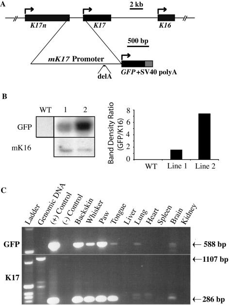
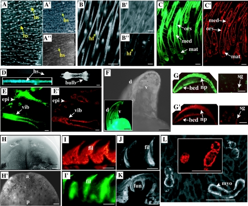
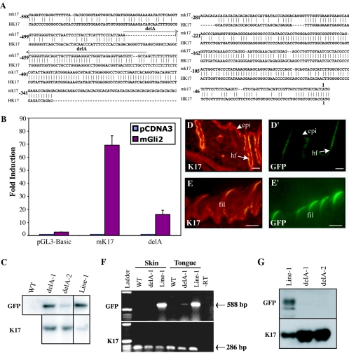
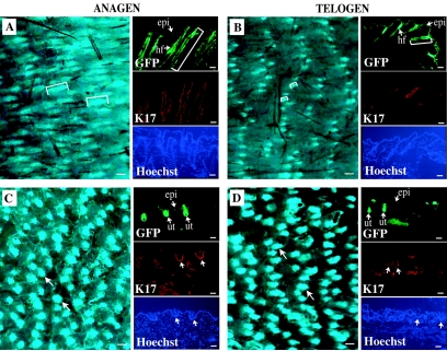
Keratin genes afford, given their large number (>50) and differential regulation, a unique opportunity to study the mechanisms underlying specification and differentiation in epithelia of higher metazoans. Moreover, the small size and regulation in cis of many keratin genes enable the use of their regulatory sequence to achieve targeted gene expression in mice. Here we show that 2 kilobases of 5' upstream region from the mouse keratin 17 gene (mK17) confers expression of green fluorescent protein (GFP) in major epithelial appendages of transgenic mice. Like that of mK17, onset of [mK17 5']-GFP reporter expression coincides with the appearance of ectoderm-derived epithelial appendages during embryonic development. In adult mice, [mK17 5']-GFP is appropriately regulated within hair, nail, glands, and oral papilla. Tracking of GFP fluorescence allows for the visualization of growth cycle-related changes in hair follicles, and the defects engendered by the hairless mutation, in live skin tissue. Deletion of an internal 48-bp interval, which encompasses a Gli-responsive element, from this promoter results in loss of GFP fluorescence in most appendages in vivo, suggesting that sonic hedgehog participates in K17 regulation. The compact mK17 gene promoter provides a novel tool for appendage-preferred gene expression and manipulation in transgenic mice.


Figures

FIG. 1.
FIG. 1.
Production and characterization of [mK17 5′]-GFP transgenic mouse lines. (A) Top, genomic context in which mK17 is located (data from reference 67). Bottom, structure of the [mK17 5′]-GFP transgene. The entire intergenic sequence between the telomeric mK17n and mK17 was subcloned upstream from the enhanced GFP coding sequence. A simian virus 40 poly(A) signal is used at the 3′end of the construct. (B) Southern blotting-based comparison of transgene copy numbers in [mK17 5′]-GFP lines 1 and 2. The single-copy mK16 is used as a reference. Quantitation was performed by densitometry. WT, wild type. (C) RT-PCR-based survey comparing GFP and K17 mRNA expression in tissues harvested from [mK17 5′]-GFP transgenic mice (line 1). “Ladder” refers to a 1-kb DNA ladder. (+) Control refers to the transgene construct. (−) Control refers to omission of the reverse transcription step. Two micrograms of total RNA was used in the reverse transcription step for each tissue. The expected amplicon products are as follows: GFP mRNA, 588 bp; K17 mRNA, 286 bp; K17 gene (reflecting genomic DNA contamination), 1,107 bp.
FIG. 2.
FIG. 2.
Analysis of GFP expression in live adult skin tissue and in tissue sections. Three-month-old adult mice were used. (A to A") Fluorescence imaging of the ear surface, shown at low magnification, in adult transgenic mice from lines 1 (A) and 2 (A"). Arrows point to protruding hair shafts (hs). A′, bright-field image for frame A. Bars, 500 μm. (B to B") Series similar to panels A to A" (with the exception that panel B" is a nontransgenic control) shown at higher magnification. Arrowheads point to signal located beneath the skin surface, reflecting hair follicles (hf). Bars, 300 μm. (C and C′) Distribution of GFP fluorescence (C) and K17 antigens (C′) in consecutive tissue sections from back skin (line 1). mat, hair matrix; med, medulla of the hair shaft; ors, outer root sheath. Bars, 50 μm. (D) Visualization of GFP fluorescence in a plucked vibrissa (vib) follicle (D) and in a section thereof (inset). Bar, 100 μm. (E and E′) Comparison of the distribution of GFP (E) and K17 (E′) in a newborn whisker pad (line 1). epi, epidermis. Bars, 100 μm. (F) Live fluorescence imaging of nail (low magnification) from an adult transgenic mouse in lines 1 (F) and 2 (inset). d, dorsal side; v, ventral side. Bars, 500 μm. (G and G′) Comparison of GFP (G) and K17 (G′) in an adult digit tip, emphasizing nail and sweat gland histology (line 1). np, nail plate; sg, sweat gland. Bars, 100 μm. (H) Bright-field image of the dorsal surface of the tongue (low magnification) from an adult transgenic mouse (line 1). (H′) Corresponding live fluorescence imaging. a, anterior; p, posterior. Bars, 500 μm. (I to L′) Analysis of tissue sections prepared from a tongue sample from an adult transgenic mouse in line 1. (I and I′) Comparison of K17 (I) and GFP (I′) in sections from dorsal tongue epithelium. (J and K) GFP fluorescence in filiform (fil) and fungiform (fun) papillae. Bars, 50 μm. (L) Comparison of the distributions of GFP (L) and K17 (inset) in glandular tissue embedded within the tongue muscle. Signal is restricted to the myoepithelial layer (myo), as expected. Bars, 50 μm.
FIG. 3.
FIG. 3.
Analysis of GFP expression in transgenic mouse embryos. All images correspond to GFP-based fluorescence seen in transgenic mouse embryos, unless stated otherwise. (A) Live imaging of an E16.5 embryo shown at low magnification. Inset, bright-field image. Bars, 1 mm. (B and B′) Comparison of the distribution of GFP fluorescence (B) and K17 antigens (B′) in consecutive tissue sections prepared from the back of an E15.5 embryo. A single placode is shown. “p” refers to the thin periderm layer covering the embryo. Bars, 50 μM. (C to L) Temporal series of live GFP fluorescence data collected from the trunk surface (C to G) and eye region (H to L) in embryos ranging from E12.5 to E16.5. Embryo age is indicated at the bottom left. In panels E to G, arrows and arrowheads depict primary and secondary placodes, respectively. In panel L, in which the eyelid is completely fused, arrows indicate sites of future eyelashes. Bars, 500 μm.
FIG. 4.
FIG. 4.
A short internal sequence confers high responsiveness to Gli2 in transfected cells and appendageal expression in vivo. All images shown correspond to GFP-based fluorescence seen in transgenic mouse embryos, unless stated otherwise. (A) Nucleotide sequence alignment (ConSite, http://mordor.cgb.ki.se/cgi-bin/CONSITE/consite/) for the proximal ∼500 bp of 5′upstream sequence from the mouse (mK17) and human K17 (hK17) genes. Vertical lines between the two sequences depict identity. The segment deleted between bp −461 and −414 in mK17 (delA) is identified by a line. (B) Luciferase reporter assays in 308 mouse keratinocytes cotransfected with Gli 2 and either [mK17 5′]-luciferase, [mK17 delA 5′]-luciferase, or pGL3-Basic (empty vector control). Luciferase activity for each construct is expressed as fold induction over the activity registered when cotransfected with pCDNA3. Error bars indicate standard errors of the means. (C) Southern blotting-based comparison of transgene copy number in [mK17 delA 5′]-GFP lines 1 and 2 with that of the parental [mK17 5′]-GFP line 1. The single-copy mK17 is used as a reference. WT, wild-type genomic DNA. (D to E′) Comparison of the distribution of K17 (D and E) and GFP (D′ and E′) fluorescence in consecutive tissue sections prepared from back skin (D and D′) and dorsal tongue epithelium (E and E′). epi, epidermis; hf, hair follicles; fil, filiform papillae. Bars, 50 μm. (F) RT-PCR-based survey comparing GFP mRNA expression in skin and tongue from wild-type (control), [mK17 5′]-GFP (line 1), and [mK17 delA 5′]-GFP (line DelA-1) transgenic mice. “Ladder,” 1-kb DNA ladder. −RT, omission of the reverse transcription step. Expected amplicon products: GFP mRNA, 588 bp; K17 mRNA, 286 bp. (G) Detection, by Western immunoblotting, of GFP (top) and K17 (bottom) antigens in total protein extracts prepared from hair clippings of [mK17 5′]-GFP (“Line-1”), and [mK17 delA 5′]-GFP (“delA-1 and delA-2”) transgenic mice.
FIG. 5.
FIG. 5.
Monitoring of progression through the hair cycle and its defects in live transgenic mouse skin tissue. (A and B) [mK17 5′]-GFP transgenic mice (line 1) at P33 and P24, respectively, at which times hair follicles are in the anagen (A) and telogen (B) phases of their cycle. (C and D) P33 (C) and P24 (D) mice homozygous for the hairless allele as well as transgenic for [mK17 5′]-GFP (line 1). Panels A, B, C, and D are fluorescence recordings of live back skin tissue. Brackets depict the length of hair follicles based on GFP fluorescence. Bars, 200 μm. The small panels shown to the right of each main panel show data gathered from consecutive sections prepared from the same back skin tissues. Labeling is indicated in their lower left corners. Hoechst staining was performed to localize individual nuclei in the preparations stained for K17. Bars, 50 μm. The arrows in panels C and D point to utricles (ut). epi, epidermis; hf, hair follicles.

References

    1. Andl, T., S. T. Reddy, T. Gaddapara, and S. E. Millar. 2002. WNT signals are required for the initiation of hair follicle development. Dev. Cell 2:643-653. - PubMed
    1. Bernot, K. M., P. A. Coulombe, and K. M. McGowan. 2002. Keratin 16 expression defines a subset of epithelial cells during skin morphogenesis and the hair cycle. J. Investig. Dermatol. 119:1137-1149. - PubMed
    1. Brakebusch, C., R. Grose, F. Quondamatteo, A. Ramirez, J. L. Jorcano, A. Pirro, M. Svensson, R. Herken, T. Sasaki, R. Timpl, S. Werner, and R. Fassler. 2000. Skin and hair follicle integrity is crucially dependent on beta 1 integrin expression on keratinocytes. EMBO J. 19:3990-4003. - PMC - PubMed
    1. Brembeck, F. H., and A. K. Rustgi. 2000. The tissue-dependent keratin 19 gene transcription is regulated by GKLF/KLF4 and Sp1. J. Biol. Chem. 275:28230-28239. - PubMed
    1. Bruen, K. J., C. A. Campbell, W. G. Schooler, S. deSerres, B. A. Cairns, C. S. Hultman, A. A. Meyer, and S. H. Randell. 2004. Real-time monitoring of keratin 5 expression during burn re-epithelialization. J. Surg. Res. 120:12-20. - PubMed

Publication types

MeSH terms

LinkOut - more resources