. 2005 Aug;25(16):7278-88.
doi: 10.1128/MCB.25.16.7278-7288.2005.

Neurexophilin 3 is highly localized in cortical and cerebellar regions and is functionally important for sensorimotor gating and motor coordination

Affiliations

Neurexophilin 3 is highly localized in cortical and cerebellar regions and is functionally important for sensorimotor gating and motor coordination

Vassilios Beglopoulos et al. Mol Cell Biol. 2005 Aug.

Abstract

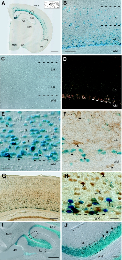
Neurexophilin 3 (Nxph3) is a specific ligand of synaptic alpha-neurexins that are essential for efficient neurotransmitter release. Previous biochemical work demonstrated that Nxph3 interacts with an extracellular domain of alpha-neurexins in a tight complex; however, no information is available on the localization or functional role of Nxph3 in the brain. Here, we generated lacZ reporter gene knock-in mice to investigate the distribution of Nxph3 at the single-cell level and Nxph3 knockout mice to examine its functional importance. Nxph3 expression was restricted mostly to subplate-derived neurons in cortical layer 6b, granule cells in the vestibulocerebellum, and Cajal-Retzius cells during development. Colabeling experiments demonstrated that neurons expressing Nxph3 do not belong to a uniform cell type. Morphological analyses and systematic behavioral testing of knockout mice revealed no anatomical defects but uncovered remarkable functional abnormalities in sensory information processing and motor coordination, evident by increased startle response, reduced prepulse inhibition, and poor rotarod performance. Since Nxph3-deficient mice behaved normally while performing a number of other tasks, our data suggest an important role for Nxph3 as a locally and temporally regulated neuropeptide-like molecule, presumably acting in a complex with alpha-neurexins in select neuronal circuits.


Figures

FIG. 1.
FIG. 1.
Generation of Nxph3-LacZ reporter gene knock-in mice. (A) Knock-in strategy. ATG, start codon; Ex, coding exon; *, stop codon; NeoR, neomycin resistance gene; F, Flag epitope; I, internal ribosomal entry site; LacZ, β-galactosidase coding sequence; 3′ UTR, 3′ untranslated region; TK, thymidine kinase; triangles, loxP sites; gray arrows, FRT sites; 5′ and 3′ probes, outside Southern probes. Abbreviations for restriction enzymes are as follows: AI, AgeI; BHI, BamHI; BII, BglII; ERI, EcoRI; NI, NcoI; and PI, PstI. (B) COS-7 cells transfected with the Nxph3 targeting sequence show enrichment of staining over nuclei as predicted (arrows). Untransfected cells (asterisks) show no staining. (C) Immunoblot analysis of COS-7 cells transfected with the Nxph 3 targeting sequence (lane 1), an untagged Nxph3 expressionplasmid (lane 2), or mock-transfected (lane 3). Blots were probed with an antibody to the Flag epitope (upper panel), identifying the Nxph3-Flag fusion protein as predicted from the results shown in panel A. A peptide antiserum (lower panel) directed against the 10 C-terminal amino acid residues detects expression of the untagged Nxph3 (lane 2), but fails to recognize the Nxph3-Flag fusion protein (lane 1). (D) Southern blot analysis of PstI/AgeI-digested genomic DNA of wild-type (lanes 1 and 2), heterozygous (lanes 3 to 6), and homozygous (lanes 7 and 8) knock-in mice probed with the 5′ outside probe. (E) Short genomic PCRs used for genotyping wild-type (lanes 1, 600-bp band), knock-in (lanes 1, 400-bp band), and knockout alleles (lanes 3) and Cre transgene (lanes 2) in various combinations. (F and G) Position of oligonucleotide primers (F) and results (G) of an RT-PCR strategy to demonstrate expression of wild-type (lanes 1) and Flag epitope-tagged (lanes 2) Nxph3 mRNA in the brain. (H) Overview of a parasagittal section (see inset) from a 20-day-old knock-in mouse processed for β-galactosidase histochemistry, resulting in a blue reaction product where Nxph3 is expressed. Brst, brainstem; Cb, cerebellum; Co, colliculi; Cp, caudate putamen; Cx, cortex; H, hippocampal formation; Hy, hypothalamus; Ob, olfactory bulb; rsCx, retrosplenial cortex; S, subiculum; Th, thalamus. Bar, 800 μm.
FIG. 2.
FIG. 2.
Nxph3 expression is highly localized in cortical and cerebellar regions. (A) Overview picture of a coronal section (see inset) from an adult Nxph3 knock-in mouse. Prominent β-galactosidase staining is seen over deep layers of isocortical areas (e.g., visual cortex, V1&2), in the subiculum and part of the amygdala (Amy, basolateral nucleus). CA1, CA1 region of the hippocampus; DG, dentate gyrus; GeN, geniculate nucleus; MaN, mammilary nucleus; Pir, piriform cortex. Bar, 800 μm. (B) Differential interference contrast picture of the area boxed in panel A. β-Galactosidase-positive cells are enriched in layer 6 (L.6) along the border to the white matter (WM). (C) Control section from a wild-type mouse processed for β-galactosidase histochemistry. (D) Dark-field picture of an in situ hybridization experiment visualizing Nxph3 on a cortical section from an adult wild-type mouse. Bar, 150 μm (for panels B to D). (E) Overall morphology of β-galactosidase-stained cells in the cortex include horizontally orientedfusiform neurons (arrows) and medium-sized pyramidal neurons (arrowheads). Asterisks indicate neurons without Nxph3 expression. Bar, 30 μm. (F) Double-staining experiment using β-galactosidase histochemistry and antibodies against glutamate decarboxylase, GAD67. (G and H) Partial colabeling of Nxph3 with tbr1 shown at low (G) and high (H) magnification (arrows). Note that more neurons in layer 6 are Tbr1-positive (arrowheads). Bar, 30 μm. (I) Parasagittal section through parts of the cerebellar vermis from 20-day-old Nxph3 knock-in mice. Lo 10, uvula; Lo 9, nodule; Med, medial cerebellar nucleus; 4V, fourth ventricle. Bar, 200 μm. (J) Higher magnification of the area boxed in panel I demonstrates that Nxph3 is expressed by cells of the granule cell layer but appears to spare Purkinje neurons (arrows). Ml, molecular layer; Pcl, Purkinje cell layer; Gl, granule cell layer; WM, white matter. Bar, 40 μm.
FIG. 3.
FIG. 3.
Nxph3 expression in cortical brain regions during development. (A) Northern blots of brains from wild-type mice at postnatal day 1 (P1), 6 (P6), 10 (P10), and 14 (P14) and at about 6 weeks (adult). RNA samples were probed for Nxph3, and β-actin for input control. (B to E) β-Galactosidase-stained sections (pictures converted to gray scale) through the cerebral cortex from Nxph3 knock-in mice at the indicated ages. Arrowheads point to transiently expressing cells in the marginal zone, vertical arrows mark a population of subplate neurons which develops into mature layer 6 neurons after the first week, and horizontal arrows identify a few scattered neurons in layer 5 that appear last. CP, cortical plate; SP, subplate. Bars, 20 μm (B), 50 μm (C), and 100 μm (E, for panels D and E).
FIG. 4.
FIG. 4.
Hippocampal Cajal-Retzius cells transiently expressing Nxph3. (A to D) Coronal sections through the dentate gyrus (DG) and hippocampus (H) of Nxph3 knock-in mice at the indicated ages. β-Galactosidase staining shows Nxph3-expressing cells mostly in the outer half of the molecular layer (ML) during the first postnatal week that gradually disappear afterwards. GCL, granule cell layer of the dentate gyrus. Bar in panel D (for panels A to D), 150 μm. (E and F) Double-staining of the dentate gyrus from a 7-day-old knock-in mouse with an antibody to reelin (immunofluorescence) (E) and with β-galactosidase histochemistry (differential-interference contrast) (F). The extracellular matrix protein reelin is distributed predominantly around β-Gal-positive cells in the molecular layer (ML; arrows), whereas no reelin staining can be seen over the granule cell layer (GCL). (G and H) Similar experiment using antibodies against calretinin, also showing colocalization with Nxph3-expressing cells (arrows). Bars, 30 μm (E and F) and 80 μm (G and H).
FIG. 5.
FIG. 5.
Generation of Nxph3 knockout mice reveals no effect of Nxph3 on brain structure. (A) Diagram depicting the strategy of generating Nxph3 knockout mice. Primer pairs for genotyping knock-in and knockout mice are indicated by the half-arrows (for genotyping samples, see Fig. 1E). Abbreviations are as defined in the legend to Fig. 1A. (B) Northern blots of wild-type, Nxph3-lacZ knock-in, and Nxph3 knockout mouse brains incubated with Nxph3 probe and β-actin as the loading control. The shift to a higher-molecular-weight mRNA species in knock-in mice reflects the cotranscription of theIRES-lacZ cassette with Nxph3. (C) Neurexophilins were enriched from the brains of wild-type, Nxph3 knockout (KO), and Nxph1 and Nxph3 double knockout mice (Nxph1 and 3 DKO) on immobilized α-latrotoxin by virtue of their tight binding to α-neurexins. The immunoblot demonstrates the precipitation of α-neurexins (lower panel) but the complete absence of Nxph1 and Nxph3 in double knockout brains (upper panel). In Nxph3 knockout mice, the remaining band is due to the cross-reactivity with Nxph1 (18, 26), which is still present in Nxph3 null mutants. (D and E) Overview pictures of Nissl-stained parasagittal sections from brains of adult wild-type and Nxph3 knockout mice. Brst, brainstem; Cb, cerebellum; Co, colliculi; Cp, caudate putamen; Cx, cortex; H, hippocampal formation; Hy, hypothalamus; Ob, olfactory bulb; Th, thalamus. Bar, 900 μm. (F to I) Immunohistochemistry using antibodies against synapsin (F and G), and vesicular glutamate transporter VGlu1 (H and I) shows punctate staining patterns in the neuropil of both wild-type (F and H) and null mutant brains (G and I). Bar in panel I (for panels F to I), 20 μm.
FIG. 6.
FIG. 6.
Impaired sensorimotor gating in Nxph3 knockout mice. (A) Amplitudes of the acoustic startle response were significantly increased in Nxph3 knockout mice (KO; n = 13) compared to littermate WT controls (n = 12). (B) Prepulse inhibition of the startle response was measured at increasing intensities of the warning tone (prepulse). Compared to WT littermate animals (n = 12), Nxph3-deficient mice (Nxph3 KO; n = 13) display a significantly reduced inhibition by the prepulse at all prepulse intensities tested. Data are expressed as means ± SEM.
FIG. 7.
FIG. 7.
Motor coordination defects in Nxph3 mutants. (A) The ability for motor coordination and balance of WT (n = 12) and Nxph3 knockout mice (Nxph3 KO; n = 17) was tested with a rotarod apparatus. During accelerating training sessions and trials with constant speed (trials at 16, 24, and 32 rpm), knockout animals exhibited significantly shorter latencies to fall of the rotating drum. n.s., not significant. (B) Measurements of swimming speed during the Morris water maze task demonstrated that Nxph3 knockout mice (n = 13) do not suffer from an overall impaired locomotor ability but can outperform even their littermate controls (n = 11). Data are expressed as means ± SEM.

References

    1. Arber, S. 2004. Subplate neurons: bridging the gap to function in the cortex. Trends Neurosci. 27:111-113. - PubMed
    1. Carlson, S., and J. F. Willott. 1998. Caudal pontine reticular formation of C57BL/6J mice: responses to startle stimuli, inhibition by tones, and plasticity. J. Neurophysiol. 79:2603-2614. - PubMed
    1. Carter, R. J., L. A. Lione, T. Humby, L. Mangiarini, A. Mahal, G. P. Bates, S. B. Dunnett, and A. J. Morton. 1999. Characterization of progressive motor deficits in mice transgenic for the human Huntington's disease mutation. J. Neurosci. 19:3248-3257. - PMC - PubMed
    1. Dean, C., F. G. Scholl, J. Choih, S. DeMaria, J. Berger, E. Isacoff, and P. Scheiffele. 2003. Neurexin mediates the assembly of presynaptic terminals. Nat. Neurosci. 6:708-716. - PMC - PubMed
    1. Del Rio, J. A., B. Heimrich, H. Super, V. Borrell, M. Frotscher, and E. Soriano. 1996. Differential survival of Cajal-Retzius cells in organotypic cultures of hippocampus and neocortex. J. Neurosci. 16:6896-6907. - PMC - PubMed

Publication types

MeSH terms