. 2005 Jul 29;33(13):4265-75.
doi: 10.1093/nar/gki731. Print 2005.

An atypical RNA pseudoknot stimulator and an upstream attenuation signal for -1 ribosomal frameshifting of SARS coronavirus

Affiliations

An atypical RNA pseudoknot stimulator and an upstream attenuation signal for -1 ribosomal frameshifting of SARS coronavirus

Mei-Chi Su et al. Nucleic Acids Res. .

Abstract

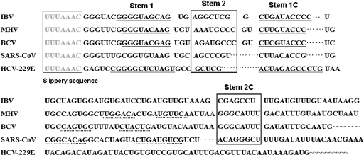
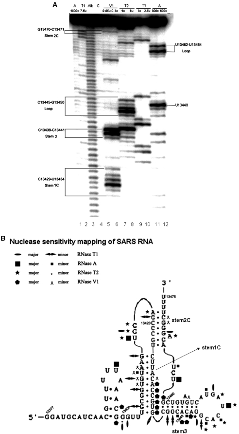
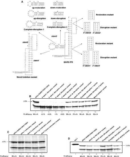
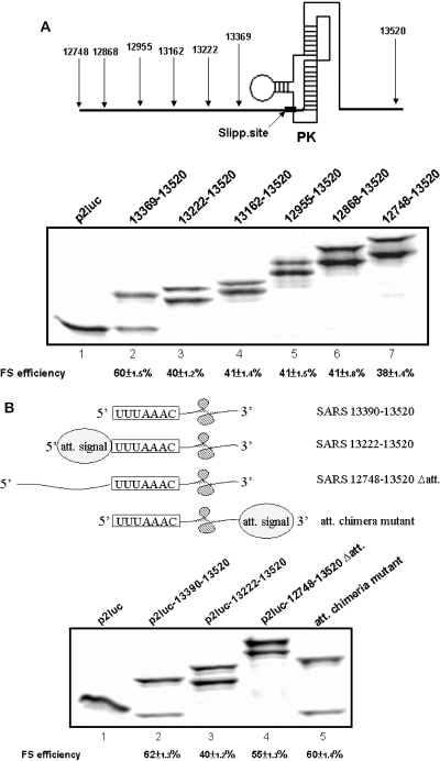
The -1 ribosomal frameshifting requires the existence of an in cis RNA slippery sequence and is promoted by a downstream stimulator RNA. An atypical RNA pseudoknot with an extra stem formed by complementary sequences within loop 2 of an H-type pseudoknot is characterized in the severe acute respiratory syndrome coronavirus (SARS CoV) genome. This pseudoknot can serve as an efficient stimulator for -1 frameshifting in vitro. Mutational analysis of the extra stem suggests frameshift efficiency can be modulated via manipulation of the secondary structure within the loop 2 of an infectious bronchitis virus-type pseudoknot. More importantly, an upstream RNA sequence separated by a linker 5' to the slippery site is also identified to be capable of modulating the -1 frameshift efficiency. RNA sequence containing this attenuation element can downregulate -1 frameshifting promoted by an atypical pseudoknot of SARS CoV and two other pseudoknot stimulators. Furthermore, frameshift efficiency can be reduced to half in the presence of the attenuation signal in vivo. Therefore, this in cis RNA attenuator represents a novel negative determinant of general importance for the regulation of -1 frameshift efficiency, and is thus a potential antiviral target.


Figures

Figure 1
Figure 1
Characterization of the in cis RNA elements involved in −1 frameshifting for SARS CoV. Sequence alignment for a set of related coronaviruses. The slippery site is boxed and typed in gray, whereas the complementary base pairing counterpart of stem regions 1 and 2 (S1-S1C and S2-S2C) of stimulator are underlined and boxed by solid line, respectively. The potential base pairing scheme for the third stem (S3) is underlined by dashed line. The S2C region for HCV 229E is not shown as it appears in further downstream region.
Figure 2
Figure 2
The SARS13 369–13 520 RNA contains an atypical H-type pseudoknot. (A) Electrophoretic analysis of probing data confirms the existence of S3. The enzymatic cleavage result was resolved in a 10% sequencing gel with the first two lanes representing pyrimidines and guanine assignment markers, respectively. The third and fourth wells are alkaline hydrolysis ladder and control, respectively. The concentration of the probes used and the assignment of residues are all shown on top of the gel directly. (B) Summary of probing data supports the predicted S3 for an atypical H-type pseudoknot within the stimulator RNA. Extent of cleavage for each probe is defined as major or minor cut as indicated by the symbols.
Figure 3
Figure 3
Role of base pairing formation in S3 may be different from those in S1 to S2 for the promotion of −1 frameshifting. (A) Illustration of mutant constructs for the manipulation of base pairing scheme. (B) Results of 12% SDS–PAGE analysis of frameshift efficiency for constructs of different base pairing scheme disruption mutants (as indicated in the top). (C) Results of 12% SDS–PAGE analysis of frameshift efficiency for different stem restoration constructs (as indicated in the top). (D) Results of 12% SDS–PAGE analysis of frameshift efficiency for different stem 3 constructs (as indicated in the top) using capped mRNA templates. The calculated efficiency is shown in the bottom of the gel and the bands of shifted product are arrowed by −1FS.
Figure 4
Figure 4
An RNA attenuation signal is characterized upstream of the slippery site. (A) Results of 12% SDS–PAGE analysis of −1 frameshifting of different upstream extension constructs with the calculated efficiency shown in the bottom of the gel. The lower and higher bands within each well belong to the shifted and the non-shifted products, respectively. The schematic diagram on top of the gel shows the relative position of the stimulator pseudoknot, the slippery site (filled box) and the extended upstream and downstream viral sequences for the constructs under analysis. (B) The attenuation signal need to work in cis and its orientation to the slippery site is crucial for its downregulation activity. Cartoons are used to illustrate the relative orientation of the attenuation signal, the slippery site and the stimulator within the inserts under analysis. The identity of insert within each reporter construct and the calculated frameshift efficiency are shown in top and bottom of the gel, respectively.
Figure 5
Figure 5
The SARS13 222–13 368 viral RNA possesses most of the attenuation activity in vitro. (A) Changing amino acids identity encoded by SARS13 222–13 368 RNA cannot abolish attenuation activity. The RNA sequences and the amino acids of polypeptide encoded by the reading frameshifted mutant are shown. The three inserted nucleotides are underlined and the encoded amino acids that remain unchanged are typed in bold face. (B) The most stable secondary structure of residue 13 222–13 368 RNA predicted by RNAstructure version 4.11 (27).
Figure 6
Figure 6
The general attenuation signal embedded in SARS13 222–13 368 RNA is a novel negative determinant for −1 frameshifting. (A) The attenuation signal also downregulates −1 frameshifting promoted by other pseudoknot stimulators. In this study, the Prl-SV40 reporter was used with the insertion of minimal IBV or MMTV pseudoknot containing or lacking the upstream attenuation signal as indicated on top of the gel. The calculated frameshift efficiency is shown in the bottom of the gel. The predicted secondary structure of the minimal IBV and MMTV pseudoknot are shown in the bottom as indicated and the slippery site is underlined. (B) Results of 12% SDS–PAGE analysis of frameshift efficiency for capped mRNA templates. The Xef1 represents Xenopus elongation factor 1α protein and is used as a protein marker control. The calculated efficiency is shown in the bottom of the gel and the bands of shifted product are arrowed by −1FS in both (A) and (B). (C) The attenuation activity of SARS13 222–13 368 RNA also functions in vivo. Comparison of frameshift efficiency for reporter constructs in the presence or absence of attenuation signal. The data are calculated from the results of dual-luciferase assay in vitro (lane 1) and in vivo (lane 2), respectively. The plotted values are the mean value calculated from three independent experiments with the error bars representing SD.

References

    1. Farabaugh P.J. Programmed translational frameshifting. Microbiol. Rev. 1996;60:103–134. - PMC - PubMed
    1. Gesteland R.F., Atkins J.F. Recoding: dynamic reprogramming of translation. Annu. Rev. Biochem. 1996;65:741–768. - PubMed
    1. Harger J.W., Meskauskas A., Dinman J.D. An ‘integrated model’ of programmed ribosomal frameshifting. Trends Biochem. Sci. 2002;27:448–454. - PubMed
    1. Namy O., Rousset J.P., Napthine S., Brierley I. Reprogrammed genetic decoding in cellular gene expression. Mol. Cell. 2004;13:157–168. - PubMed
    1. Dinman J.D., Ruiz-Echevarria M.J., Peltz S.W. Translating old drugs into new treatments: ribosomal frameshifting as a target for antiviral agents. Trends Biotechnol. 1998;16:190–196. - PMC - PubMed

Publication types

MeSH terms

Substances