. 2005 Oct;146(4):487-97.
doi: 10.1038/sj.bjp.0706363.

Reduced hyperpolarization in endothelial cells of rabbit aortic valve following chronic nitroglycerine administration

Affiliations

Reduced hyperpolarization in endothelial cells of rabbit aortic valve following chronic nitroglycerine administration

Nobuyoshi Kusama et al. Br J Pharmacol. 2005 Oct.

Abstract

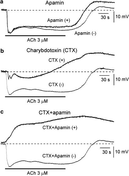
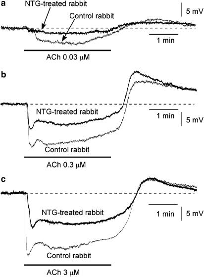
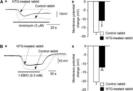
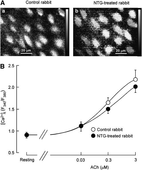
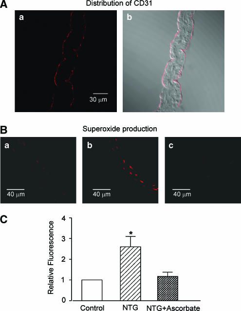
This study was undertaken to determine whether long-term in vivo administration of nitroglycerine (NTG) downregulates the hyperpolarization induced by acetylcholine (ACh) in aortic valve endothelial cells (AVECs) of the rabbit and, if so, whether antioxidant agents can normalize this downregulated hyperpolarization. ACh (0.03-3 microM) induced a hyperpolarization through activations of both apamin- and charybdotoxin-sensitive Ca2+-activated K+ channels (K(Ca)) in rabbit AVECs. The intermediate-conductance K(Ca) channel (IK(Ca)) activator 1-ethyl-2-benzimidazolinone (1-EBIO, 0.3 mM) induced a hyperpolarization of the same magnitude as ACh (3 microM). The ACh-induced hyperpolarization was significantly weaker, although the ACh-induced [Ca2+]i increase was unchanged, in NTG-treated rabbits (versus NTG-untreated control rabbits). The hyperpolarization induced by 1-EBIO was also weaker in NTG-treated rabbits. The reduced ACh-induced hyperpolarization seen in NTG-treated rabbits was not modified by in vitro application of the superoxide scavengers Mn-TBAP, tiron or ascorbate, but it was normalized when ascorbate was coadministered with NTG in vivo. Superoxide production within the endothelial cell (estimated by ethidium fluorescence) was increased in NTG-treated rabbits and this increased production was normalized by in vivo coadministration of ascorbate with the NTG. It is suggested that long-term in vivo administration of NTG downregulates the ACh-induced hyperpolarization in rabbit AVECs, possibly through chronic actions mediated by superoxide.


Figures

Figure 1
Figure 1
Effects of apamin, charybdotoxin (CTX) and apamin+CTX on acetylcholine (ACh)-induced membrane potential changes in aortic valve endothelial cells (AVECs) from NTG-untreated control rabbits. (a) Effects of apamin (0.1 μM). ACh (3 μM)-induced membrane potential changes before (‘Apamin (−)') and during (‘Apamin (+)') application of apamin. Resting membrane potentials (RMPs) were −51.5 mV and −51.0 mV before and during application of apamin, respectively. (b) Effects of CTX (0.1 μM). Before (‘CTX(−)') and during (‘CTX(+)') application of CTX. The RMPs were −51.5 and −50.6 mV before and during application of CTX, respectively. (c) Effects of CTX (0.1 μM)+apamin (0.1 μM). Before (‘CTX+Apamin (−)') and during (‘CTX+Apamin (+)') application of CTX+apamin. The RMPs were −51.5 and −50.0 mV before and during application of CTX+apamin, respectively. After recording the ACh-induced response, followed by a 25-min interval, one of the toxins or a combination was applied for 5 min, and ACh was then applied in the presence of the toxin(s). The two overlaying records shown in a given panel (a–c) were obtained from a single endothelial cell.
Figure 2
Figure 2
Effects of chronic in vivo administration of nitroglycerine (NTG) on ACh-induced membrane potential changes in AVECs. (a) Effects of 0.03 μM ACh on membrane potential in an AVEC from an NTG-untreated control rabbit (‘Control rabbit') or an NTG-treated rabbit (‘NTG-treated rabbit'). The RMP was −51.4 mV in Control rabbit and this being −51.5 mV in NTG-treated rabbit. (b) Effects of 0.3 μM ACh (as in a). The RMP was −51.2 mV in Control rabbit and this being −51.3 mV in NTG-treated rabbit. (c) Effects of 3 μM ACh (as in a). The RMP was −51.3 mV in Control rabbit and this being −51.4 mV in NTG-treated rabbit. The concentration-dependent effects of ACh shown in (a)–(c) were obtained from a single endothelial cell in each type of rabbit.
Figure 3
Figure 3
Effects of chronic in vivo NTG administration on the hyperpolarization induced by the Ca2+-ionophore ionomycin or the IKCa agonist 1-EBIO in AVECs. (A) Effects of ionomycin (3 μM) on membrane potential in AVECs from NTG-untreated control rabbits (‘Control rabbit') and NTG-treated rabbits (‘NTG-treated rabbit'). (a) Actual traces. The RMP was −52.3 mV in Control rabbit and this being −52.2 mV in NTG-treated rabbit. (b) Summary of the effect of ionomycin on membrane potential. Mean of data from four different preparations (from four different animals) with s.e.m. (B) Effects of 1-EBIO (0.3 mM) (as in A). (a) Actual traces. The RMP was −52.5 mV in Control rabbit and this being −52.3 mV in NTG-treated rabbit. (b) Summary of the effect of 1-EBIO. Mean of data from six to seven different preparations (from six to seven different animals) with s.e.m. *P<0.05, **P<0.01 versus ‘Control rabbit'.
Figure 4
Figure 4
Effects of in vivo chronic application of NTG on ACh-induced increase in intracellular concentration of Ca2+ ([Ca2+]i) in AVECs. (A) Increase in [Ca2+]i in AVECs at 3 min after application of ACh (3 μM). The [Ca2+]i is expressed as the Fura-2 ratio (F340/F380). (a) Control rabbit; (b) NTG-treated rabbit. (B) Concentration-dependent effects of ACh on [Ca2+]i in AVECs (from rabbits with or without NTG treatment). Each concentration of ACh was intermittently applied for 3 min with a 10-min interval. The maximum response induced by ACh at a given concentration was plotted. Mean of data from five different preparations (from five different animals) with s.e.m.
Figure 5
Figure 5
Superoxide production in aortic valves from rabbits treated in vivo, or not treated, with NTG or NTG+ascorbate. (A) Fluorescence image of antibody against the endothelial cell marker CD31(Aa) in a cross-section of an aortic valve, together with its bright field image (Ab). (B) Superoxide production in cross-sections of aortic valves. (Ba) NTG-untreated control rabbit; (Bb) NTG-treated rabbit; (Bc) NTG+ascorbate-treated rabbit. Superoxide was detected by dihydroethydium fluorescence. The images shown in (Ba)–(Bc) were taken under identical conditions. Similar results were obtained from four other preparations (from four different animals). (C) Summary of the effect of in vivo NTG administration on superoxide production. Relative fluorescence intensity was calculated using the aortic valve section from NTG-untreated control rabbit (Control) as standard. Mean of data from four different sections (from four different animals) with s.e.m. *P<0.05 versus ‘Control'.

Comment in

Similar articles

Cited by

References

    1. BÉNY J. Electrical coupling between smooth muscle cells and endothelial cells in pig coronary arteries. Pflügers Arch. 1997;433:364–367. - PubMed
    1. BÉNY J., PACICCA C. Bidirectional electrical communication between smooth muscle and endothelial cells in the pig coronary artery. Am. J. Physiol. 1994;266:H1465–H1472. - PubMed
    1. BERKENBOOM G., FONTAINE D., UNGER P., BALDASSARRE S., PREUMONT N., FONTAINE J. Absence of nitrate tolerance after long-term treatment with ramipril: an endothelium-dependent mechanism. J. Cardiovasc. Pharmacol. 1999;34:547–553. - PubMed
    1. BRAKEMEIER S., EICHLER I., KNORR A., FASSHEBER T., KOHLER R., HOYER J. Modulation of Ca2+-activated K+ channel in renal artery endothelium in situ by nitric oxide and reactive oxygen species. Kidney Int. 2003;64:199–207. - PubMed
    1. BRUNET P., BÉNY J. Substance P and bradykinin hyperpolarize pig coronary artery endothelial cells in primary culture. Blood Vessels. 1989;26:228–234. - PubMed

Publication types

MeSH terms